
Multi-perspective interventional imaging using a single imaging system

a multi-perspective, imaging system technology, applied in the field of imaging, can solve the problems of increasing complexity of interventions, affecting patient positioning, and gantry rotation being intrusive and often prohibitive, and affecting patient positioning

Active Publication Date: 2017-03-23
GENERAL ELECTRIC CO
View PDF1 Cites 12 Cited by
  • Summary
  • Abstract
  • Description
  • Claims
  • Application Information

AI Technical Summary

Benefits of technology

The present patent describes a method and system for obtaining tomographic images of a target region in a subject. The method includes selecting tomographic angle sequences, acquiring image sequences corresponding to the selected tomographic angle sequences, deriving geometric information and visualization information, generating displacement maps based on the geometric and visualization information, transforming images based on the displacement maps to create stabilized presentation of the target region, and visualizing the transformed images on a display. The technical effects of this patent include improved accuracy and stability of target region visualization and reduced motion blur and noise in the image sequences.

Problems solved by technology

Consequently, increasingly complex interventions are being performed thereby necessitating imaging systems to provide expanded and additional functionality in order to support these complex interventions.
However, the gantry rotation is workflow intrusive and often prohibitive due to the proximity of auxiliary equipment such as an anesthesiology cart, an ultrasound imaging system, lines to the patient, and the like.
Furthermore, workflow is also hampered by patient positioning due to radial access and / or off-center anatomy / region of interest (ROI).
Consequently, temporal information that is desirable for catheter guidance or observing bolus dynamics, for example, may not be available.
Moreover, CBCT imaging is further complicated as CBCT imaging needs to be synchronized with the contrast agent injection and / or breath hold of the patient, in order to avoid a loss in image quality, for example due to patient motion.
However, navigating the catheter through complex vasculature employing the single 2D viewing plane is laborious and time consuming Consequently, procedure time, radiation dose, as well as contrast medium dose are impacted.
However, the bi-plane system is very expensive in terms of equipment and required facility.
Moreover, maneuvering the second imaging plane into position is workflow intrusive.
Procedure X-ray dose, contrast dose, and procedure duration associated with the currently available techniques also negatively impact patient outcome and procedure cost.
Other disadvantages of the presently available techniques also include exposure of clinical staff to radiation.

Method used


Image

  • Multi-perspective interventional imaging using a single imaging system
  • Multi-perspective interventional imaging using a single imaging system
  • Multi-perspective interventional imaging using a single imaging system

Examples


Embodiment Construction

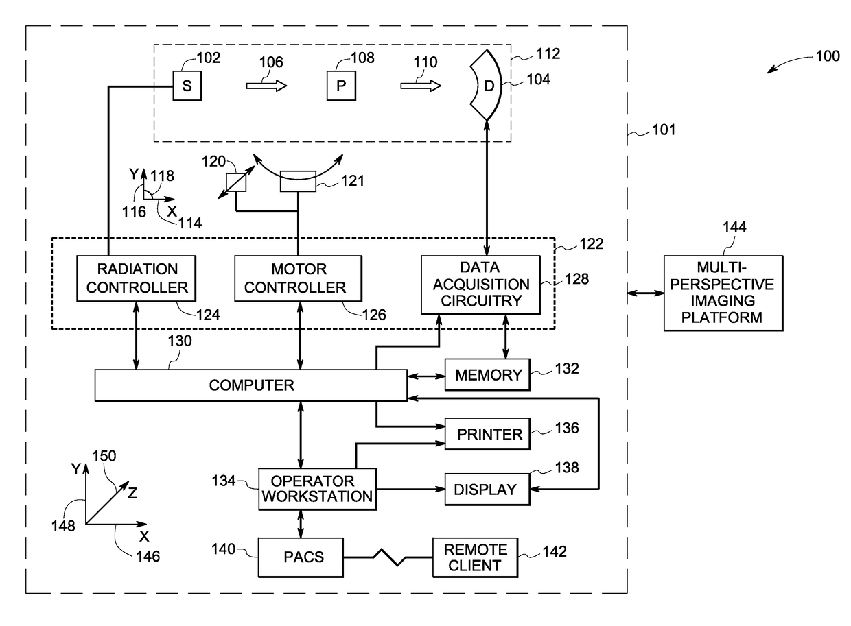
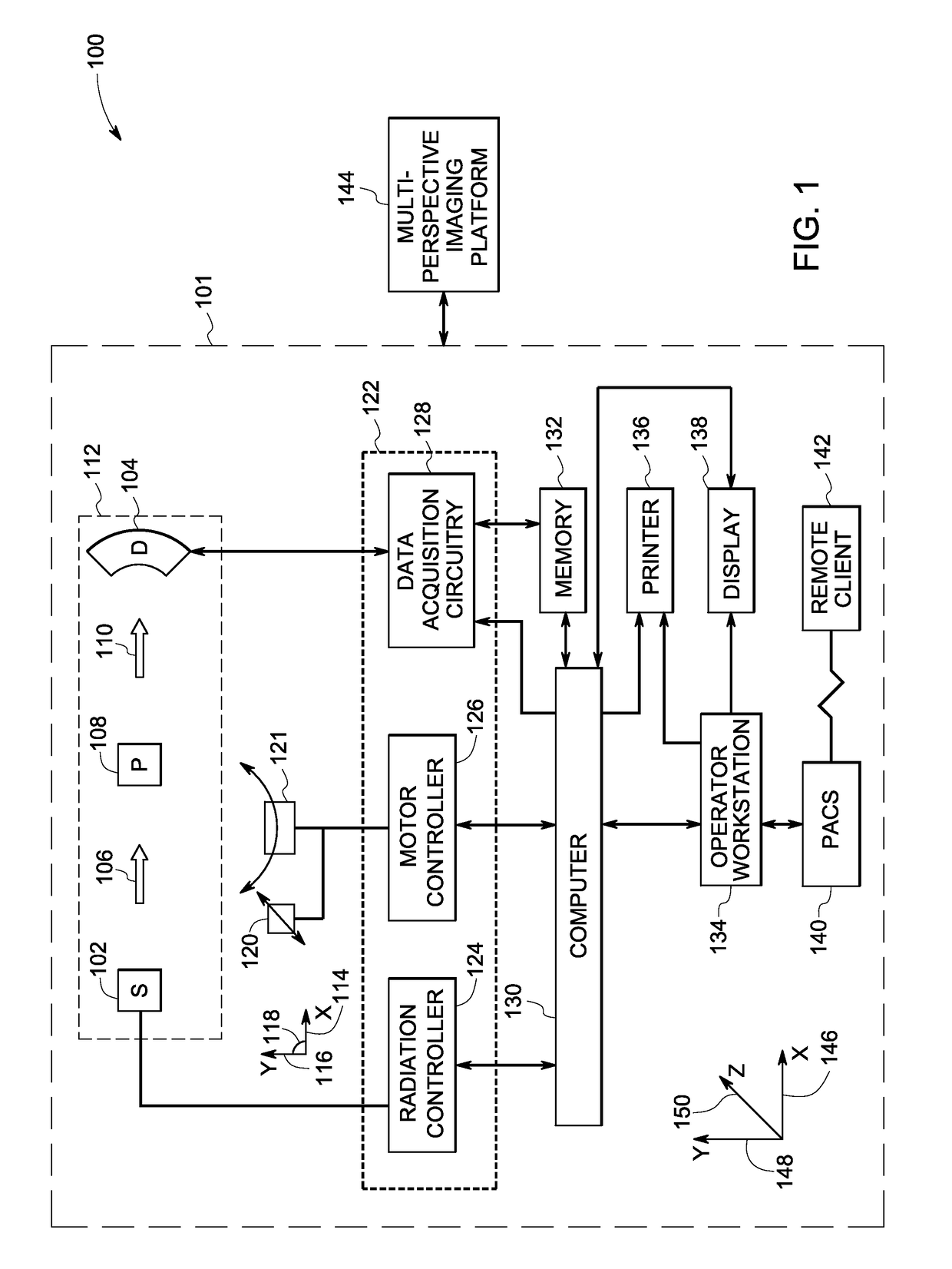
[0018]As will be described in detail hereinafter, systems and methods for multi-perspective imaging using a single imaging system, in accordance with aspects of the present specification, are presented. More particularly, the systems and methods are configured to generate one or more stabilized image sequences and present the stabilized image sequences to a user. Use of the exemplary systems and methods presented hereinafter provides enhanced visualization of structures within an imaged volume, thereby providing visualization support to the user for diagnosis and guidance support during interventional procedures.

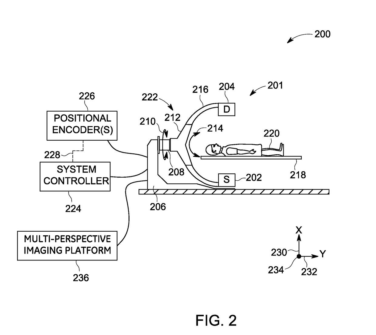
[0019]In certain 3D imaging procedures including procedures using an interventional C-arm imaging system or similar systems, it may be desirable to visualize internal structures of a patient. Aspects of the present specification utilize a C-arm imaging system to provide such images. In particular, a C-arm imaging system may be operated in an exemplary tomosynthesis acquisiti...


PUM


Abstract

A method for imaging a target region in a subject is presented. The method includes selecting one or more tomographic angle sequences, acquiring one or more image sequences corresponding to the one or more tomographic angle sequences, where each image sequence has a corresponding tomographic angle sequence, deriving geometric information corresponding to one or more structures of interest in at least one of the image sequences, identifying visualization information, generating one or more displacement maps based on the geometric information, the visualization information, at least a subset of at least one of the one or more tomographic angle sequences, or combinations thereof, transforming at least a subset of images in the one or more image sequences based on corresponding displacement maps to create one or more transformed / stabilized image sequences, and visualizing on a display the one or more transformed / stabilized image sequences to provide a stabilized presentation of the target region.

Description

BACKGROUND[0001]Embodiments of the present specification relate to imaging, and more particularly to a system and method for multi-perspective imaging via use of a single plane interventional imaging system.[0002]Minimally invasive medical procedures are becoming increasingly important due to their success in improving patient outcome and minimizing cost. Consequently, increasingly complex interventions are being performed thereby necessitating imaging systems to provide expanded and additional functionality in order to support these complex interventions. These interventional procedures may include catheter-based techniques and / or needle-based techniques. The catheter-based interventional techniques entail navigating a catheter through vasculature of the patient to reach a target region such as a diseased region. In the needle-based techniques, a needle is guided through the anatomy to reach a target region. The needle-based techniques are typically used to deliver therapy to a can...

Claims


Application Information

Patent Timeline
Patent Type & Authority Applications(United States)
IPC IPC(8): A61B6/00A61B6/02
CPCA61B6/5211A61B6/461A61B6/025A61B6/027A61B6/4441A61B6/463A61B6/486A61B6/504G16H50/20
Inventor CLAUS, BERNHARD ERICH HERMANNLANGAN, DAVID ALLEN
Owner GENERAL ELECTRIC CO
Learn More