
Image processing method and system for analyzing a multi-channel image obtained from a biological tissue sample being stained by multiple stains

a biological tissue sample and multi-channel image technology, applied in image analysis, image enhancement, instruments, etc., can solve the problem that the immunoscore study is no longer reproducible, achieve high resolution, reduce the subjectivity of independent readers, and improve the reliability and efficiency of the fov selection process

Active Publication Date: 2017-06-01
PROVIDENCE HEALTH SYST OREGON +1
View PDF8 Cites 39 Cited by
  • Summary
  • Abstract
  • Description
  • Claims
  • Application Information

AI Technical Summary

Benefits of technology

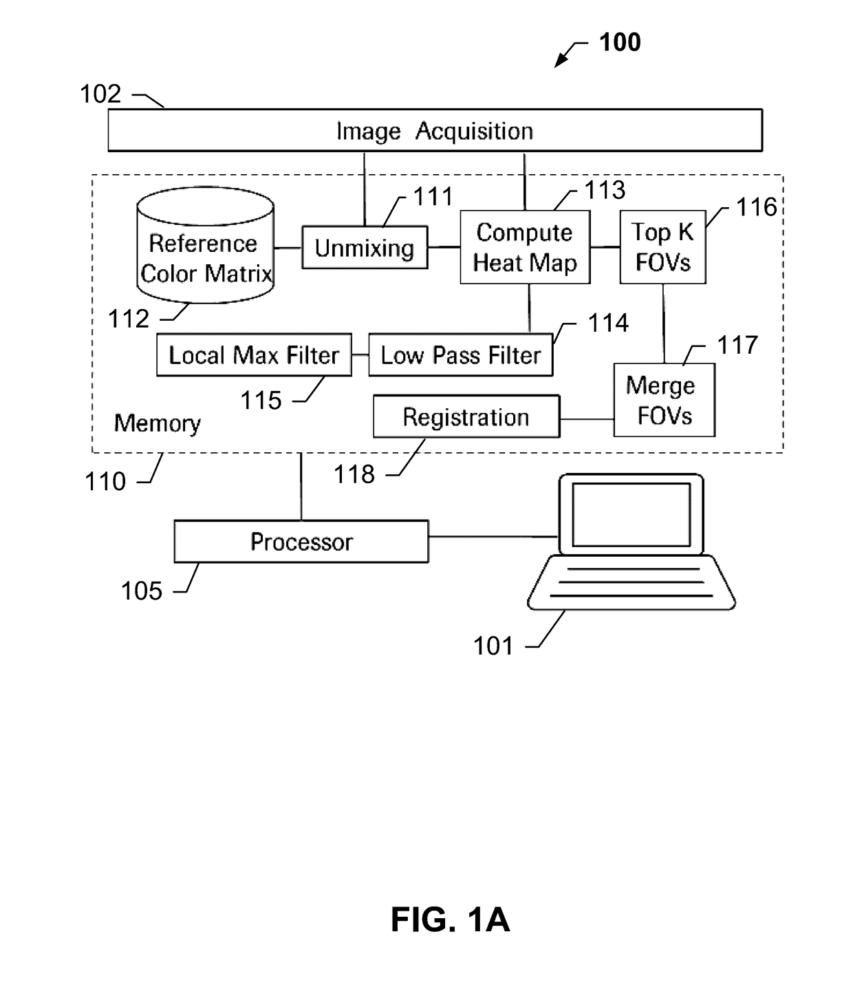
The present invention provides an automatic and reliable technique to identify fields of view in a multi-channel image without the need for manual marking by a pathologist or biologist. A spatial low pass filtering, local maximum filtering, and thresholding operations can be executed at high processing speeds, minimizing computational expense and latency time. Additionally, the invention offers a tissue mask that can be applied to the unmixed image prior or after the spatial low pass or local maximum filtering or thresholding operations and before or after the fields of view are defined. The invention uses low-resolution images to speed up the field of view selection process, reducing subjectivity and computational load. The invention includes a computer-readable medium to store computer-readable code for automated field of view selection.

Problems solved by technology

Manual selection of the FOVs and counting is highly subjective and biased to the readers, as different readers may select different FOVs to count.
Hence, an immunoscore study is no longer reproducible.

Method used


Image

  • Image processing method and system for analyzing a multi-channel image obtained from a biological tissue sample being stained by multiple stains
  • Image processing method and system for analyzing a multi-channel image obtained from a biological tissue sample being stained by multiple stains
  • Image processing method and system for analyzing a multi-channel image obtained from a biological tissue sample being stained by multiple stains

Examples


Embodiment Construction

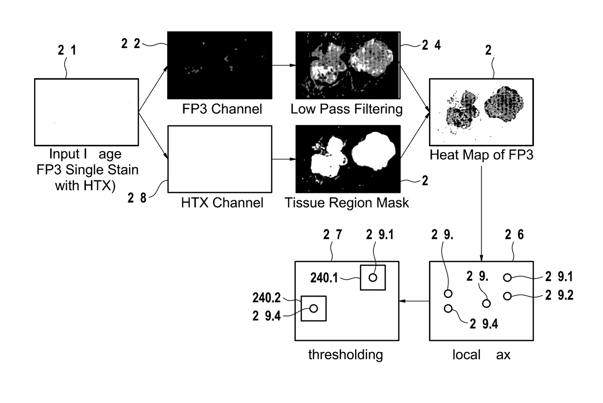
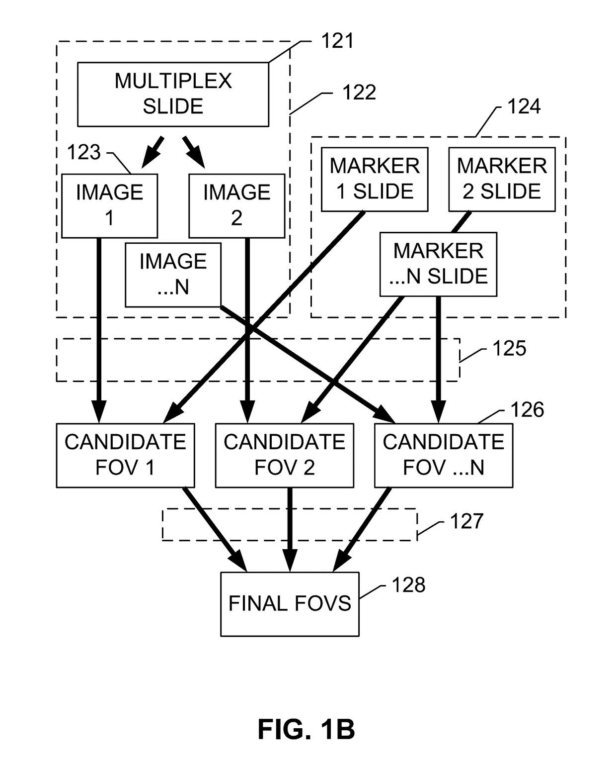
[0061]The present invention features systems and methods for automatic field of view (FOV) selection based on a density of each cell marker in a whole slide image. Operations described herein include but are not limited to reading images for individual markers from an unmixed multiplex slide or from singularly stained slides, and computing the tissue region mask from the individual marker image. A heat map of each marker may be determined by applying a low pass filter on an individual marker image channel, and selecting the top K highest intensity regions from the heat map as the candidate FOVs for each marker. The candidate FOVs from the individual marker images may then be merged together. The merging may comprise one or both of adding all of the FOVs together in the same coordinate system, or only adding the FOVs from the selected marker images, based on an input preference or choice, by first registering all the individual marker images to a common coordinate system and merging ...


PUM


Abstract

Systems and methods for automatic FOV selection in immunoscore computation that involve reading images for individual markers from an unmixed multiplex slide or single stain slides, and computing the tissue region mask from the individual marker image. The heat map of each marker is determined by applying the low pass filter on the individual marker image channel and selecting the top K highest intensity regions from the heat map as the candidate FOVs for each marker. The candidate FOVs from the individual marker images are merged together in the same coordinate system by either adding all of the FOVs together or by only adding the FOVs from the selected marker images depending on the user's choice, and registering all the individual marker images to a common coordinate system and transferring the FOVs back to the original images.

Description

CROSS-REFERENCE TO RELATED APPLICATIONS[0001]This patent application is a continuation of International Application No. PCT / EP2015 / 056770 filed Mar. 27, 2015, which claims priority to and the benefit of U.S. Provisional Application No. 62 / 005,222, filed May 30, 2014. Each of the above patent applications is incorporated herein by reference as if set forth in its entirety.BACKGROUND OF THE SUBJECT DISCLOSURE[0002]Field of the Subject Disclosure[0003]The present subject disclosure relates to imaging for medical diagnosis. More particularly, the present subject disclosure relates to automatic field of view (FOV) selection on a whole slide image.[0004]Background of the Subject Disclosure[0005]In the analysis of biological specimens such as tissue sections, blood, cell cultures and the like, biological specimens are stained with one or more combinations of stains to identify, for example, biomarkers, cells or cellular structures, and the resulting assay is viewed or imaged for further an...

Claims


Application Information

Patent Timeline
Patent Type & Authority Applications(United States)
IPC IPC(8): G06T7/00G06T7/11G06T5/00G06T5/20
CPCG06T7/0012G06T5/20G06T7/11G06T2207/30072G06T2207/10064G06T2207/30024G06T2207/10024G06T5/002G06T5/73G06T5/70
Inventor BARNES, MICHAELBIFULCO, CARLOCHEN, TINGTUBBS, ALISA
Owner PROVIDENCE HEALTH SYST OREGON
Learn More