Instrument guidance system for sinus surgery

a sinus surgery and guidance system technology, applied in the direction of instruments, image enhancement, applications, etc., can solve the problems of not providing differentiation and subdivision of individual nasal cells, not suitable for the determination of sinus natural drainage pathways, and only working with air-filled nasal cells

Active Publication Date: 2017-08-17
STRYKER EUROPEAN OPERATIONS LIMITED
View PDF6 Cites 83 Cited by
  • Summary
  • Abstract
  • Description
  • Claims
  • Application Information

AI Technical Summary

Benefits of technology

[0029]The method may further comprise that the virtual image additionally comprises visualisation elements for an enhanced 3D perception of the geometric primitive or tube shaped object.

Problems solved by technology

Disadvantages related to the method disclosed by Zein et al are that the method only works with air-filled nasal cells.
Further, it does not provide a differentiation and subdivision of individual nasal cells and the detection of natural drainage pathways of the sinuses.
It is disadvantageous that the method disclosed by Lo and de Bruijne is only applicable for segments air-filled cavities and adjusted for use to segment airways in the lung and not suitable for the determination of natural drainage pathways of the sinuses.
It is disadvantageous that only segmentation of connected cavities is possible and a manual definition of seed points for region growing algorithm is necessary.
The disadvantages are same as mentioned above for the publication of Tingelhoff et al., namely that only segmentation of connected cavities is possible and a manual definition of seed points for region growing algorithm is necessary.
A disadvantage of the method disclosed in WO 2013 / 012492 A2 is the difficulty for the user to prove the results of automatic segmentation and optimize it, especially to separate connected cells.
In addition, there is no description how to segment blocked ostia.
WO 2008 / 021702 A2 does not provide the implementation of the manual or automatic segmentation of sinus cavities
The challenge in the planning of sinus surgery, however, lies rather in a separation and identification of individual cells in particular under the absence of air.
In case of diseases such as inflammation of the sinuses or polyps, single or multiple cells are filled with tissue or fluid and the automatic segmentation methods are likely to fail due to marginal grey value differences of some cartilaginous cell walls and mucosa or pathological cell filling.
Also, the identification and labelling of separated cavities is an unsolved problem especially in the case of pathological anatomy.
Also the different quality of the 3D image data is often a problem for automatic methods, which require a high resolution of the volume image data and a normalized, or uniform grey scale density map in order to guarantee satisfying results.
Especially image data from older devices and Cone beam computed tomography (CBCT) often do not meet these conditions.
Systems known from the state of the art do not provide support for a planning scheme based on patient-specific 3D image data of the human body, particularly of the paranasal sinuses.
Therefore, up to date the planning scheme can only be manually performed on paper with the following disadvantages:Spatial incorrectnessError-prone scaling / size assignmentUsed cuboids or cylinders describe the shape of the cells sometimes inadequatelyInsufficient applicability to the surgical intervention
The perception of depth information of superimposed data in the augmented reality representation is usually also a problem, since the distance between the visualised object and the camera is often difficult to recognize.
Another challenge is the intraoperative positioning and alignment of instruments relative to planning object, particularly using the real-time endoscopic or microscopic imaging.

Method used


Image

  • Instrument guidance system for sinus surgery
  • Instrument guidance system for sinus surgery
  • Instrument guidance system for sinus surgery

Examples


Embodiment Construction

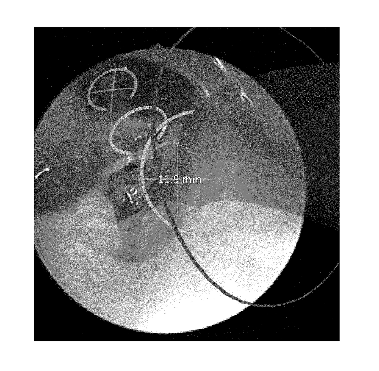
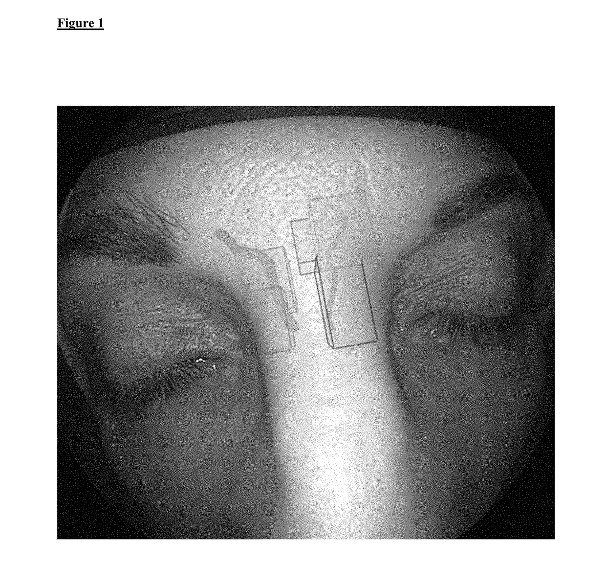
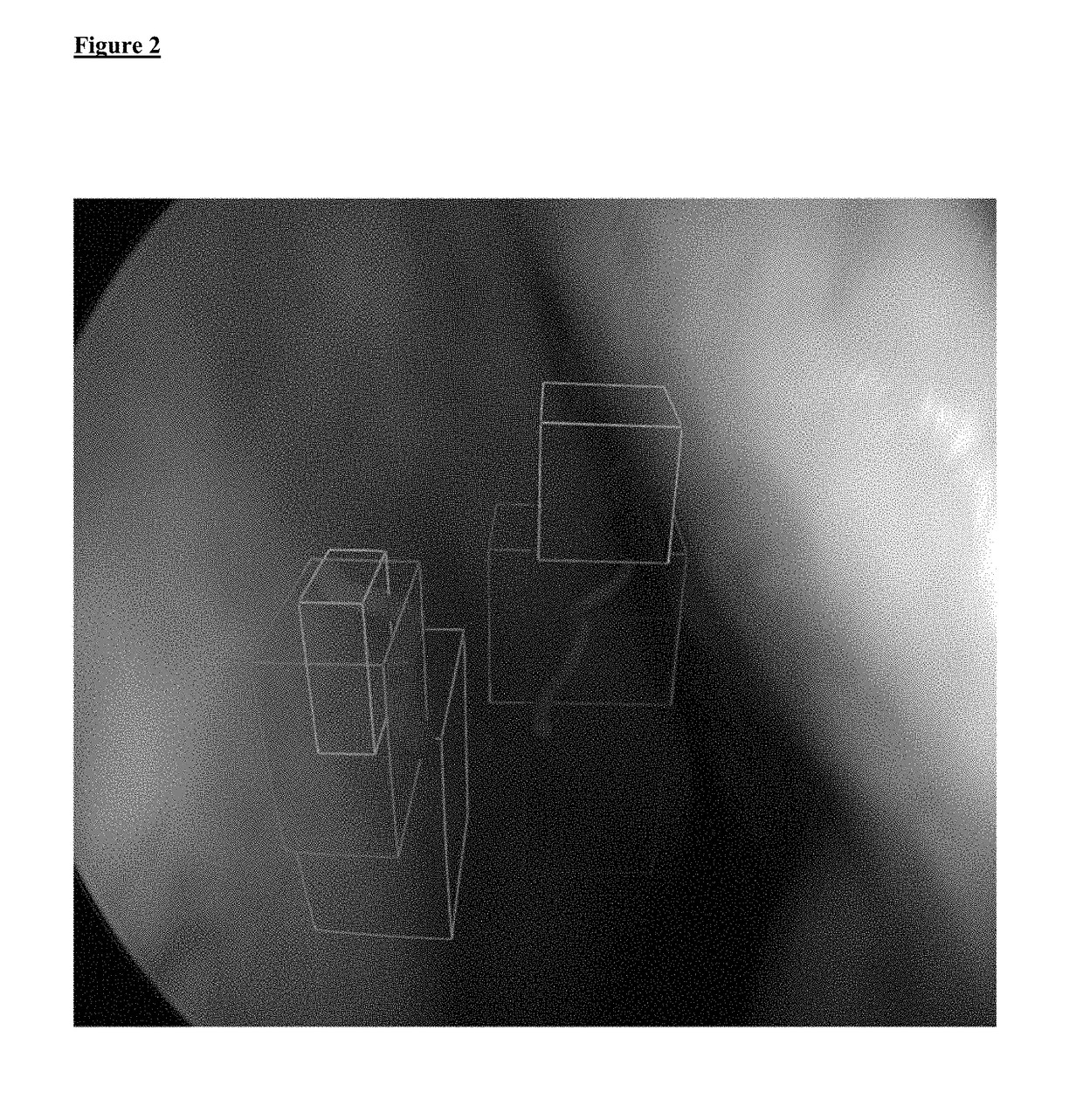
[0050]The present invention provides a fast, easy-to-use and intuitive method for generating an augmented reality image for supporting the adjustment of the position of a surgical instrument during sinus surgery. Further, the present invention provides a method for supporting computer assisted navigation and opening or reopening natural cavities and orifices of the human body, wherein the treatment of the human or animal body is not part of the instant invention. The method is based on a manual or automatic selection or segmentation of sinus cells, cavities and / or orifices of the sinuses. The method is suitable for computer assisted labelling of cavities like sinus cavities for instance in the 3-D patient image data according to the planning scheme described in the literature and taught in the training of surgeons.

[0051]It is an advantage of the subject matter of the instant invention that it is for the first time possible to assist a surgeon during sinus surgery by superimposing ge...


PUM


Abstract

A method for generating an augmented reality image for supporting the adjustment of the position of a surgical instrument during sinus surgery comprising the steps of: selecting at least one of sinus cells, cavities and orifices of the sinus in a pre-operative image by marking them with at least one geometric primitive or tube shaped object; using real-time intra-operative imaging for displaying the operating field; determining the relative positions at least between the real-time imaging of the patient and the pre-operative image data; computing a virtual image corresponding to a real-time image comprising at least one marked geometric primitive or tube shaped object based on the determined relative positions; combining the virtual image and the real-time intraoperative image for visualization in an augmented reality image.

Description

CROSS-REFERENCE TO RELATED APPLICATIONS[0001]The present application is a continuation of PCT / EP2015 / 075158 (WO 2016 / 066759) filed on Oct. 29, 2015 claiming priority of UK Patent Application No. GB 1501157.0 filed on Jan. 23, 2015 and U.S. provisional application U.S. 62 / 073,512 filed on Oct. 31, 2014. The aforementioned applications are hereby incorporated by reference in its entirety.BACKGROUND OF THE INVENTIONField of the Invention[0002]The present invention relates to a method for generating an augmented reality image for supporting the adjustment of the position of a surgical instrument during sinus surgeryBrief Description of the Related Art[0003]There are several diseases known which can cause airway narrowing or stenosis. Inflammatory conditions belong to those known diseases. While there are several methods available to improve airway narrowing, balloon dilatation may provide a gentle, uniform dilation with less airway trauma than endoscopic sinus surgery with cutting instr...

Claims


Application Information

Patent Timeline
Patent Type & Authority Applications(United States)
IPC IPC(8): A61B90/00A61B34/20G06T7/73G06T19/00G06T15/04G06K9/00A61B17/24A61B34/10
CPCA61B90/37G06T2207/10088A61B34/20A61B34/10G06T19/006G06T15/04G06K9/0014G06T7/73A61B2090/365A61B2034/105A61B2034/107A61B2034/2051A61B2034/2055G06T2207/30204G06T2207/20221G06T2207/10081A61B17/24A61B90/36G06T2210/41G06T7/10G06V20/695
Inventor KOSMECKI, BARTOSZOEZBEK, CHRISTOPHERWINNE, CHRISTIAN
Owner STRYKER EUROPEAN OPERATIONS LIMITED
Why Eureka
Try Eureka