Operating method for a medical imaging apparatus

Inactive Publication Date: 2017-11-02
SIEMENS HEALTHCARE GMBH
View PDF16 Cites 0 Cited by
  • Summary
  • Abstract
  • Description
  • Claims
  • Application Information

AI Technical Summary

Benefits of technology

The present invention provides an operating method for a medical imaging apparatus that allows for easy and coordinated changes to parameters for imaging. Each routine examination step and variant thereof is assigned an identification code for quick and efficient recognition. Users can create variants of routine examination steps by changing the step, making it easier to develop improvements or optimize the examination. This method reduces errors and allows for efficient use of existing routines.

Problems solved by technology

Improvements memorialized in this way are consequently poorly documented and often barely still comprehensible for the respective user himself or herself after some time.
Such changes to the routine examination steps are generally not accessible at all and / or often also not comprehensible to other users.

Method used


Image

  • Operating method for a medical imaging apparatus
  • Operating method for a medical imaging apparatus
  • Operating method for a medical imaging apparatus

Examples


Embodiment Construction

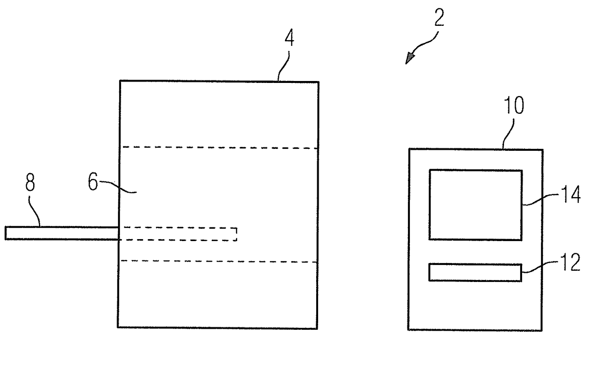
[0018]FIG. 1 shows a diagnostic magnetic resonance (MR) apparatus 2 with an MR data acquisition scanner 4. The scanner has a tunnel-shaped inner space 6, in which a patient supported on a patient couch 8 will be positioned for imaging. The scanner contains, as is known, gradient coils for spatially encoding MR signals and radio-frequency coils for excitation of nuclear spins and for receipt of the resulting MR signals, as well as further hardware components. The scanner 4 is controlled by a control computer 10. The control computer 10 includes a system processor and an image processor with associated data memories, as well as a user interface, which is connected to the system processor and the image processor, and control software. The control computer 10 additionally includes a pulse sequence controller and a gradient pulse shape generator. The user interface includes an input unit 12 and a monitor 14. The positioning of the patient and the operation of the gradient and radio-frequ...


PUM


Abstract

An operating method for a medical imaging apparatus includes an imaging program stored in a memory, with which parameters for controlling image data generation will be predetermined. The imaging program has a number of routine examination steps. At least one variant, which is likewise stored in the memory and which is able to be selected by a user instead of the routine examination step for image data generation, exists at least for one of the routine examination steps.

Description

BACKGROUND OF THE INVENTIONField of the Invention[0001]The present invention relates to an operating method for a medical imaging apparatus with an imaging program stored in a memory, with which parameters for controlling generation of imaging data will be predetermined, wherein the imaging program includes a number of routine examination steps.Description of the Prior Art[0002]A medical imaging apparatus is an apparatus for acquiring, processing, evaluating and / or storing imaging information in the form of image data. Acoustic methods such as ultrasound (US), emission methods such as Emission Computed Tomography (ECT) and Positron Emission Tomography (PET), optical methods, radiological methods such as x-ray tomography and computed Tomography (CT) can be used to acquire the imaging information for example. The acquisition can also be done by Magnetic Resonance Tomography (MR or MRT) or by combined methods. The medical imaging apparatus can deliver 2-dimensional (2D) or multidimensi...

Claims


Application Information

Patent Timeline
IPC IPC(8): G01R33/54A61B5/00A61B5/055G06F19/00G16H10/60
CPCG01R33/54A61B5/0555A61B5/742A61B2576/00G06F19/322G01R33/543G06F19/3406G16H40/63G16H10/60G01R33/546A61B5/055
Inventor SCHOR, STEFAN
Owner SIEMENS HEALTHCARE GMBH
Why Eureka
Try Eureka