Method and apparatus for providing information to a patient during a medical imaging examination that indicates the residual examination time for the examination

Inactive Publication Date: 2018-03-22
SIEMENS HEALTHCARE GMBH
View PDF9 Cites 0 Cited by
  • Summary
  • Abstract
  • Description
  • Claims
  • Application Information

AI Technical Summary

Benefits of technology

[0033]The different positions for display signals along the progress bar are preferably color-coded so that the signal color of a display signal alone can be used to draw conclusions with respect to the output information. In addition, a color profile of a number of display signals on the progress bar can be used to depict a time profile of the medical imaging examination for the patient. For example, the start of the progress bar and hence the start of the medical imaging examination can be given a red color. The middle of the progress bar, and hence the middle of the medical imaging examination, can then be given a yellow and/or orange color. The end of the progress bar, and hence the end of the medical imaging examination, can be given a green color. Hence, the patient can use the display symbol displayed to acquire the current status of the medical imaging examination in terms of time in a simple and quick manner.
[0034]In another embodiment of the invention, the optical output information is at least one light signal. A light signal means a li

Problems solved by technology

Patients frequently find it difficult to estimate the remaining examination time correctly during a medical imaging examination.
This can cause a patient to experience u

Method used


Image

  • Method and apparatus for providing information to a patient during a medical imaging examination that indicates the residual examination time for the examination
  • Method and apparatus for providing information to a patient during a medical imaging examination that indicates the residual examination time for the examination
  • Method and apparatus for providing information to a patient during a medical imaging examination that indicates the residual examination time for the examination

Examples


Example

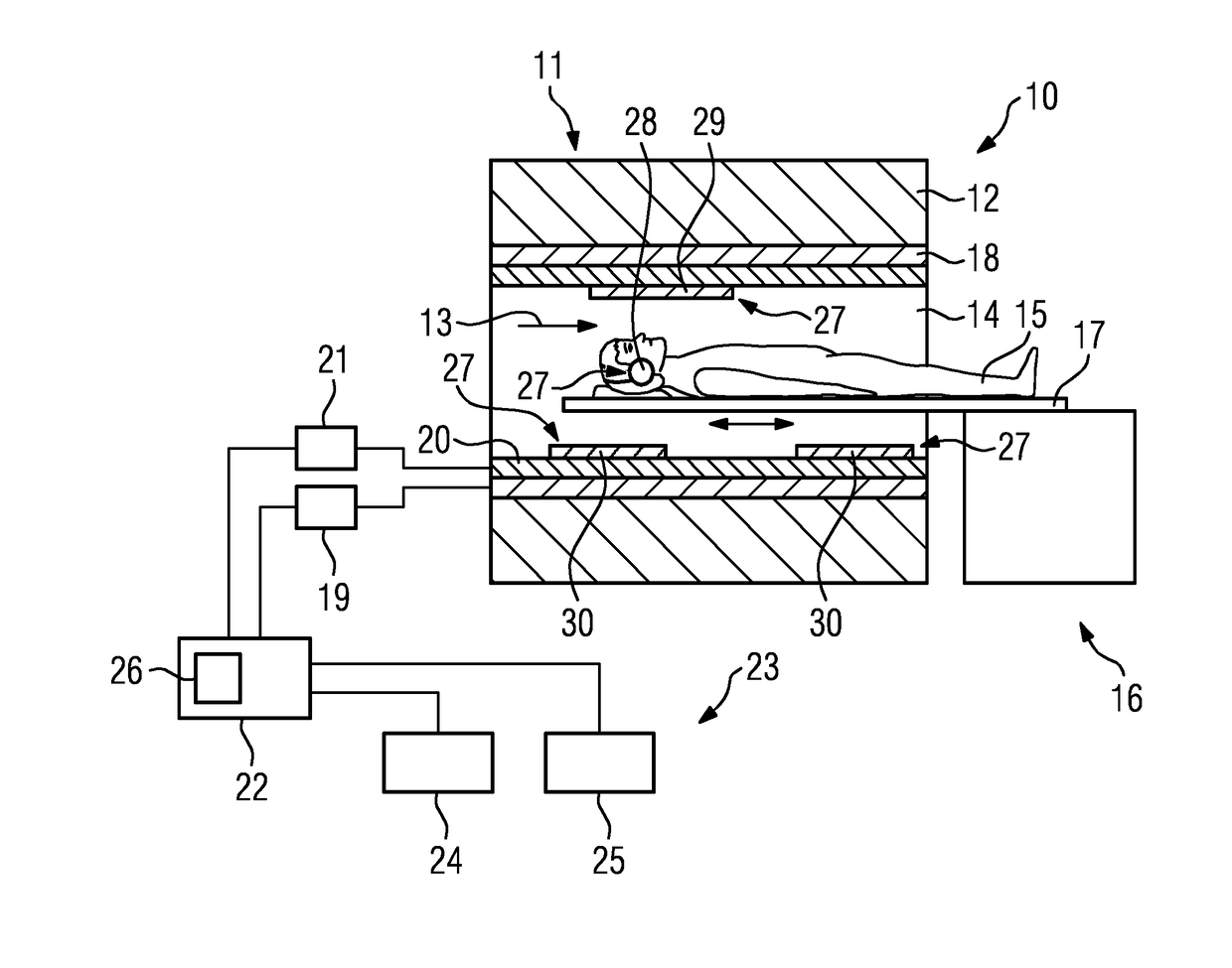
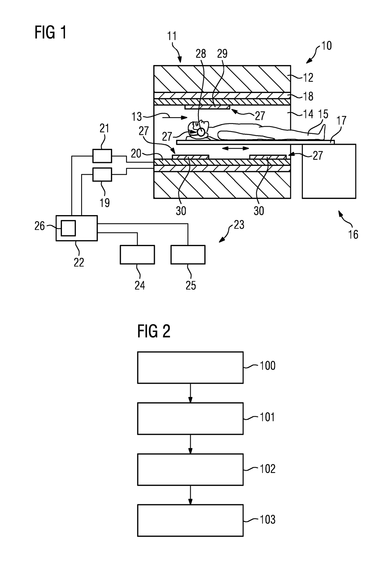
[0045]FIG. 1 is a schematic depiction of a medical imaging apparatus. In the present exemplary embodiment, the medical imaging apparatus is a magnetic resonance apparatus 10, so the present invention will be explained with the example of the magnetic resonance apparatus 10. However, the present invention is not restricted to an embodiment of the medical imaging apparatus as a magnetic resonance apparatus 10 and further embodiments of the medical imaging apparatus are conceivable, such as an embodiment of the medical imaging apparatus as a computed tomography apparatus or as a PET apparatus.

[0046]The magnetic resonance apparatus 10 has a scanner 11 with a superconducting basic field magnet 12 that generates a strong, constant, basic magnetic field 13. The scanner 11 also has a patient-receiving region 14 for receiving a patient 15. In the present exemplary embodiment, the patient-receiving region 14 has a cylindrical shape and is surrounded circumferentially by the scanner 11. Embodi...


PUM


Abstract

A method for providing information to a patient during a medical imaging examination, said method comprising: operating a medical imaging apparatus, while a patient is undergoing an examination with the medical imaging apparatus, during said medical imaging examination, providing a computer with time information that informs said computer as to an amount of time that has elapsed from a beginning of said medical imaging examination, from said time information, determining, in said computer, a residual examination time for the medical imaging examination that designates an ending time at which said medical imaging examination is expected to end, generating output information in said computer dependent on said residual examination time, said output information comprising humanly perceptible information formatted to inform the patient as to when said medical imaging examination is expected to end; and providing said output information in said humanly perceptible form to the patient while the patient is undergoing said medical imaging examination.

Description

BACKGROUND OF THE INVENTIONField of the Invention[0001]The present invention relates to a method, a medical imaging apparatus, and a non-transitory data storage medium for providing output information to a patient during a medical imaging examination, wherein the information indicates the residual examination time for the medical imaging examination.Description of the Prior Art[0002]Medical imaging examinations of a patient can take varying lengths of time depending on the type of the medical imaging examination and / or depending on the body region of the patient and / or depending on the medical and / or diagnostic issue in question. An examination can take from a few minutes to an hour or longer. Particularly for magnetic resonance examinations, it is usual for examinations to take an hour and longer.[0003]Patients frequently find it difficult to estimate the remaining examination time correctly during a medical imaging examination. This can cause a patient to experience uncertainty or...

Claims


Application Information

Patent Timeline
IPC IPC(8): A61B5/00G07C3/04
CPCA61B5/743A61B5/7415G07C3/04A61B90/08A61B5/055A61B5/742A61B6/032A61B6/037A61B6/46A61B2090/0807G01R33/283
Inventor NUFER, STEPHANKUERTEN, ANJASCHUBART, LEATHOENE, JENS
Owner SIEMENS HEALTHCARE GMBH
Why Eureka
Try Eureka