Methods and system for shading a two-dimensional ultrasound image

a two-dimensional ultrasound and ultrasound image technology, applied in image enhancement, instruments, ultrasonic/sonic/infrasonic image/data processing, etc., can solve the problem of increasing the difficulty of diagnosing a patient using the flat 2d image, and affecting the diagnostic accuracy of patients

Active Publication Date: 2018-11-08
GENERAL ELECTRIC CO
View PDF3 Cites 10 Cited by
  • Summary
  • Abstract
  • Description
  • Claims
  • Application Information

AI Technical Summary

Problems solved by technology

However, the dataset may include 2D scalar data (e.g., intensity values, power component values, or the like) which results in a flat 2D image that may be more difficult to interpret, thereby increasing a difficulty of diagnosing a patient using the flat 2D image.
For example, more complex body structures may be difficult to recognize via a 2D image.
As one example, 2D color Doppler images of different body structures may be especially difficult to use for diagnosis.

Method used


Image

  • Methods and system for shading a two-dimensional ultrasound image
  • Methods and system for shading a two-dimensional ultrasound image
  • Methods and system for shading a two-dimensional ultrasound image

Examples


Embodiment Construction

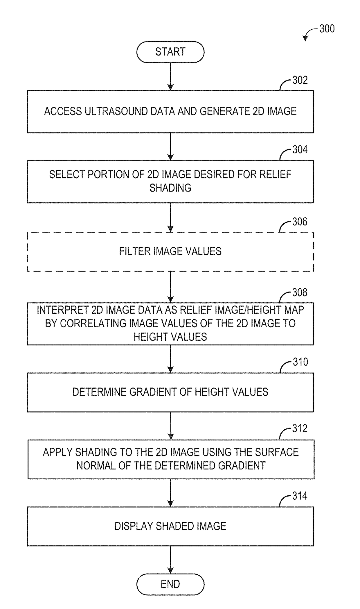
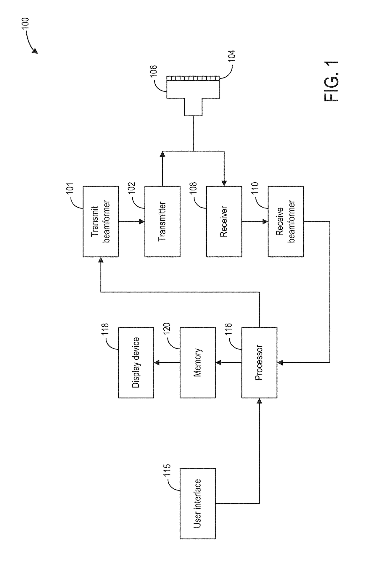
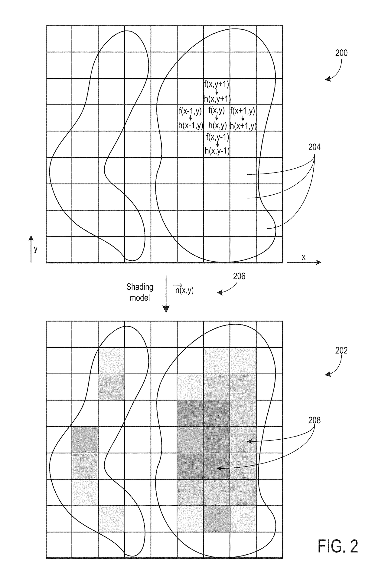
[0014]The following description relates to various embodiments of shading a 2D ultrasound image using a gradient determined from height values correlated to image values of an ultrasound imaging dataset (which may be 1D, 2D, or 3D) used to generate the 2D ultrasound image. An ultrasound system, such as the system shown in FIG. 1, may be used to acquire the ultrasound imaging dataset. A 2D image may then be generated using the acquired dataset. An example of a 2D ultrasound image generated from an imaging dataset is shown in FIG. 2. FIG. 2 also shows an example of a shaded 2D ultrasound image where the shading is applied to the 2D ultrasound image using a gradient surface normal determined from image values of the dataset that have been converted to height values. For example, as shown by the method of FIG. 3, image values, such as power or intensity values, of an acquired ultrasound imaging dataset may be converted to height values. A gradient is then determined for the converted he...


PUM


Abstract

Various methods and systems are provided for shading a 2D ultrasound image, generated from ultrasound data, using a gradient determined from scalar values of the ultrasound image data. As one example, a method includes correlating image values of a dataset acquired with an ultrasound imaging system to height values; determining a gradient of the height values; applying shading to a 2D image generated from the dataset using the determined gradient; and displaying the shaded 2D image.

Description

FIELD[0001]Embodiments of the subject matter disclosed herein relate to applying shading to a two-dimensional ultrasound image.BACKGROUND[0002]An ultrasound imaging system may be used to acquire images of a patient's anatomy. Ultrasound imaging systems may acquire a dataset which is then used to generate a 2D image that a medical professional may view and use to diagnose a patient. However, the dataset may include 2D scalar data (e.g., intensity values, power component values, or the like) which results in a flat 2D image that may be more difficult to interpret, thereby increasing a difficulty of diagnosing a patient using the flat 2D image. For example, more complex body structures may be difficult to recognize via a 2D image. As one example, 2D color Doppler images of different body structures may be especially difficult to use for diagnosis.BRIEF DESCRIPTION[0003]In one embodiment, a method comprises correlating image values of a dataset acquired with an ultrasound imaging system...

Claims


Application Information

Patent Timeline
Patent Type & Authority Applications(United States)
IPC IPC(8): G06T7/00G06T7/50A61B8/08A61B8/00
CPCG06T7/0012G06T7/50A61B8/488G06T2207/10132A61B8/5207A61B8/54A61B8/461A61B8/5215G06T11/00A61B8/06G06T15/80
Inventor SCHROECKER, GERALDBUCKTON, DANIEL JOHN
Owner GENERAL ELECTRIC CO
Why Eureka
Try Eureka