Predictive diagnostic workflow for tumors using automated dissection, next generation sequencing, and automated slide stainers

a technology of automated dissection and tumors, applied in the direction of instruments, biochemistry apparatus and processes, material analysis, etc., can solve the problems of not providing any information about the spatial relationship of mutations or how mutations are inter-related from a functional standpoin

Pending Publication Date: 2018-11-29
VENTANA MEDICAL SYST INC
View PDF0 Cites 17 Cited by
  • Summary
  • Abstract
  • Description
  • Claims
  • Application Information

AI Technical Summary

Benefits of technology

[0007]Methods are provided in which regions of a tumor sample predicted not to respond to a first therapeutic agent are excised from the sample with an automated dissection tool, mutations

Problems solved by technology

While these methods may provide a high-level view of mutations represented in the tumor, they do not provide any informa

Method used


Image

  • Predictive diagnostic workflow for tumors using automated dissection, next generation sequencing, and automated slide stainers
  • Predictive diagnostic workflow for tumors using automated dissection, next generation sequencing, and automated slide stainers
  • Predictive diagnostic workflow for tumors using automated dissection, next generation sequencing, and automated slide stainers

Examples


embodiment 7

8. The method of embodiment 7, wherein the solid tumor is a formalin-fixed, paraffin-embedded (FFPE) tissue sample, and the first sample and the additional sample(s) are microtome sections of the FFPE tissue sample.

embodiment 8

9. The method of embodiment 8, wherein first sample and the additional sample(s) are serial sections.

10. The method of any of embodiments 1-9, wherein the next generation sequencer operates on a principle selected from the group consisting of pyrosequencing, cyclic reversible termination, semiconductor sequencing technology, and phospholinked fluorescent nucleotides.

11. The method of any of embodiments 1-10, wherein the first predictive biomarker and the additional predictive biomarker(s) are selected from the group consisting of ALK, ATM, BCL2, BRAF, BRCA1, c-KIT, CAIX, CCR4, CD30, Claudin, 17p13.1, DLL3, EGFR1, estrogen receptor, EREG, ERCC1, FGF19, FGFR2b, FGFR3, FOLR1, hyaluronan, HER2 / NEU, K-ras, MGMT, MSLN, p53, MDM2, progesterone receptor, PD-L1, PDGFRB, PTEN, and thymidine phosphorylase.

12. A system comprising:[0132](a) a set of microscope slides comprising:[0133](a1) a first microscope slide having deposited thereon a first sample of a tumor, wherein the first sample is his...

embodiment 14

15. The system of embodiment 14, wherein the label automatically directs the automated slide stainer to execute the instructions on the second sample.

16. The system of embodiment 14, wherein the label generates a report for an operator of the automated slide stainer, the report instructing the manual operator to program the automated slide stainer to execute the instructions on the second sample.

17. The system of any of embodiments 12-16, wherein the next generation sequencer operates on a principle selected from the group consisting of pyrosequencing, cyclic reversible termination, semiconductor sequencing technology, and phospholinked fluorescent nucleotides.

18. The system of any of embodiments 12-17, wherein the first predictive biomarker and the additional predictive biomarker(s) are selected from the group consisting of ALK, ATM, BCL2, BRAF, BRCA1, c-KIT, CAIX, CCR4, CD30, Claudin, 17p13.1, DLL3, EGFR1, estrogen receptor, EREG, ERCC1, FGF19, FGFR2b, FGFR3, FOLR1, hyaluronan, HE...


PUM

PropertyMeasurementUnit
Fluorescenceaaaaaaaaaa

Abstract

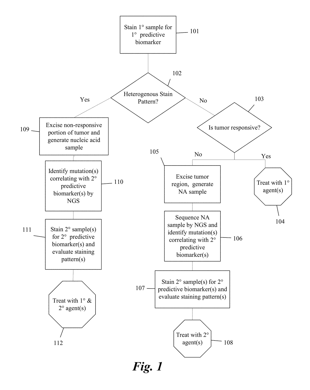
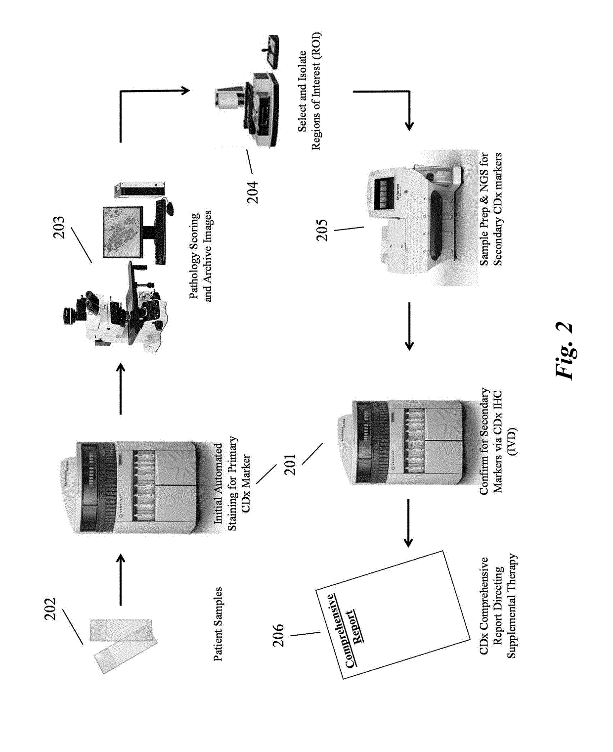
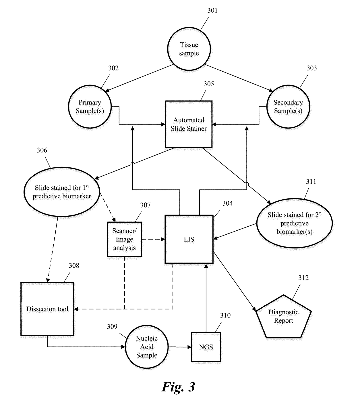
Systems and methods for selecting therapeutic agents for cancers using next generation sequencing, automated dissection, and/or automated slide stainers are disclosed. Non-responsive regions of a tumor sample having a heterogenous staining pattern for a predictive biomarker are excised using an automated dissection tool. Mutations linked to additional predictive biomarkers are identified in the excised portion of the sample by next generation sequencing. The relevance of the additional predictive biomarker(s) is confirmed by histochemical staining. Therapeutic courses may then be selected on the basis of the staining patterns of the predictive biomarkers.

Description

CROSS-REFERENCE TO RELATED APPLICATIONS[0001]This is a continuation of PCT / US2017 / 014969, filed Jan. 26, 2017, which claims the benefit of U.S. Provisional Patent Application No. 62 / 287,182, filed Jan. 26, 2016, the content of each of which is incorporated herein by reference in its entirety.BACKGROUND OF THE INVENTIONField of the Invention[0002]The invention relates to use of automated slide stainers, automated dissection tools, and / or next generation sequencers to assay predictive biomarkers and / or select therapies for cancers.Description of Related Art[0003]In the last 20 years, cancer research and treatment has undergone a sea change. Whereas the first sequencing of the human genome cost on the order of $500 million to $1 billion, next generation sequencing (NGS) strategies have shrunk that cost to less than $2000, thereby permitting large-scale and relatively inexpensive evaluation of cancer genetics. See Reuter et al. These advances have accelerated the rate at which new thera...

Claims


Application Information

Patent Timeline
IPC IPC(8): G01N1/31C12Q1/6869G01N1/28G01N35/00
CPCG01N1/312C12Q1/6869G01N1/2813G01N35/0092G01N35/00732C12Q2600/106C12Q2600/156G01N2001/282C12Q1/6806C12Q1/6841G01N1/31C12Q2535/122C12Q2537/149C12Q2537/165
Inventor GUSTAFSON, HEATHERHNATYSZYN, HARRY JAMES
Owner VENTANA MEDICAL SYST INC
Why Eureka
Try Eureka