
System for generating a synthetic 2d image with an enhanced depth of field of a biological sample

a biological sample and synthetic image technology, applied in the field of synthetic 2d image generating system with enhanced depth of field of biological sample, can solve problems such as the depth of field of the microscop

Active Publication Date: 2019-03-07
KONINKLJIJKE PHILIPS NV
View PDF6 Cites 7 Cited by
  • Summary
  • Abstract
  • Description
  • Claims
  • Application Information

AI Technical Summary

Benefits of technology

The patent describes a microscope scanner that can capture images of a biological sample at different positions. The scanner can also capture images at different depths to create a synthetic 2D image with enhanced depth of field. This means that the scanner can update the image based on new image data that is captured as it sweeps past a certain position. This technique allows for better image quality without saving every previous image, resulting in improved efficiency and accuracy in biological analysis.

Problems solved by technology

As these samples contain 3D structures and the depth of field of the microscope is limited, not all parts will be in focus.

Method used


Image

  • System for generating a synthetic 2d image with an enhanced depth of field of a biological sample
  • System for generating a synthetic 2d image with an enhanced depth of field of a biological sample
  • System for generating a synthetic 2d image with an enhanced depth of field of a biological sample

Examples


Embodiment Construction

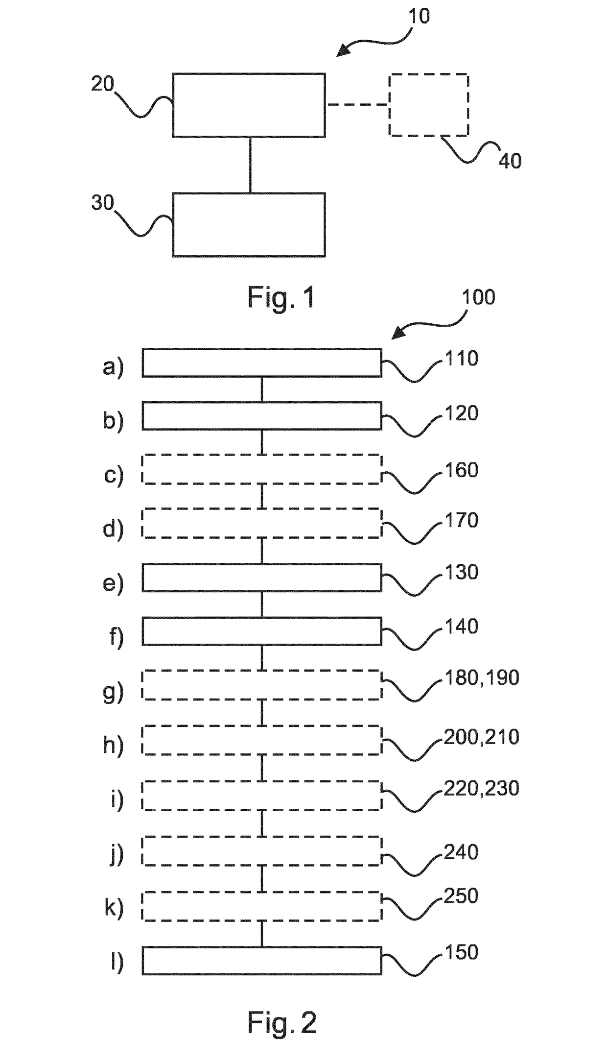
[0059]FIG. 1 shows a system 10 for generating a synthetic 2D image with an enhanced depth of field of a biological sample. The system 10 comprises: a microscope-scanner 20 and a processing unit 30. The microscope scanner 20 is configured to acquire first image data at a first lateral position of the biological sample and second image data at a second lateral position of the biological sample. The microscope scanner 20 is also configured to acquire third image data at the first lateral position and fourth image data at the second lateral position. The third image data is acquired at a depth that is different than that for the first image data and the fourth image data is acquired at a depth that is different than that for the second image data. The processing unit 30 is configured to generate first working image data for the first lateral position, the generation comprising processing the first image data and the third image data by a focus stacking algorithm. The processing unit 30 ...


PUM


Abstract

The present invention relates to a system for generating a synthetic 2D image with an enhanced depth of field of a biological sample. It is described to acquire (110) with a microscope-scanner (20) first image data at a first lateral position of the biological sample and second image data at a second lateral position of the biological sample. The microscope-scanner is used to acquire (120) third image data at the first lateral position and fourth image data at the second lateral position, wherein the third image data is acquired at a depth that is different than that for the first image data and the fourth image data is acquired at a depth that is different than that for the second image data. First working image data is generated (130) for the first lateral position, the generation comprising processing the first image data and the third image data by a focus stacking algorithm. Second working image data is generated (140) for the second lateral position, the generation comprising processing the second image data and the fourth image data by the focus stacking algorithm. The first working image data and the second working image data are combined (150), during acquisition of image data, to generate the synthetic 2D image with an enhanced depth of field of the biological sample.

Description

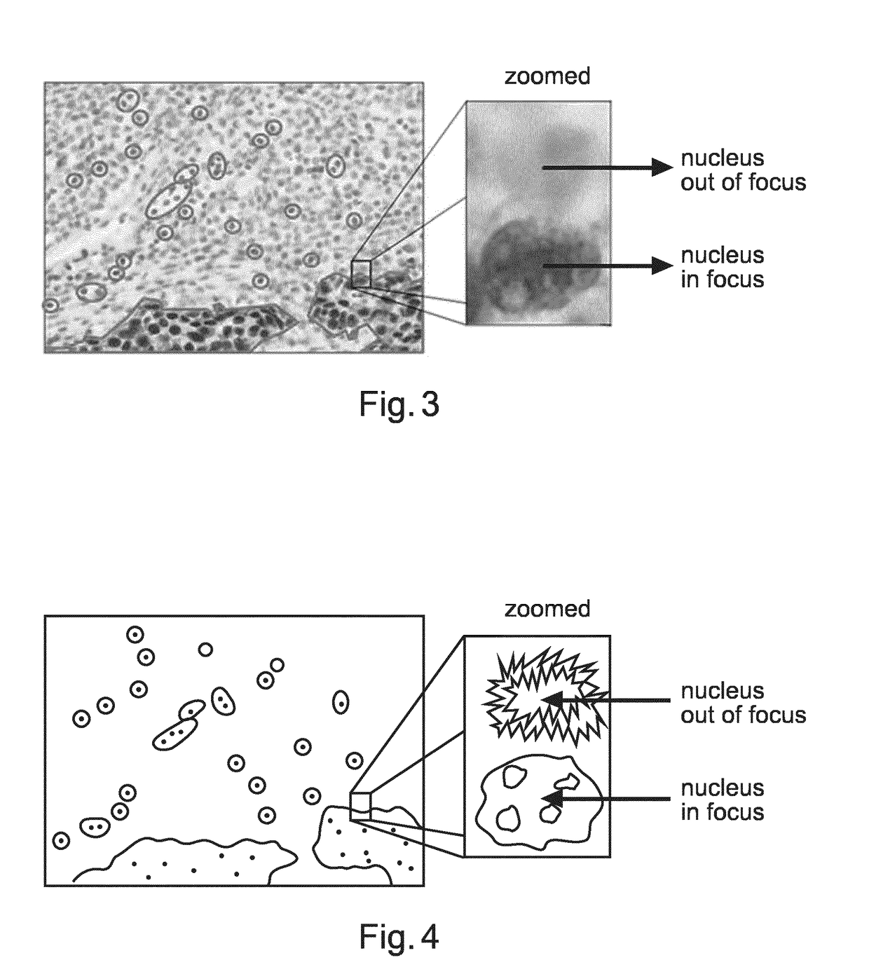
FIELD OF THE INVENTION[0001]The present invention relates to a system for generating a synthetic 2D image with an enhanced depth of field of a biological sample, to a method for generating a synthetic 2D image with an enhanced depth of field of a biological sample, as well as to a computer program element and a computer readable medium.BACKGROUND OF THE INVENTION[0002]In traditional cancer diagnosis, (histo-)pathological images of tissue samples are visually analysed by pathologists. Using a microscope, the pathologist inspects the tissue. As these samples contain 3D structures and the depth of field of the microscope is limited, not all parts will be in focus. By turning the focus knob, the pathologist is able to go through the tissue in the z-direction (i.e. depth direction). However, using a digital microscope, the tissue samples are automatically scanned. In case the tissue is scanned at a single depth, not all parts of the tissue can be in focus. Scanning the slides at multiple...

Claims


Application Information

Patent Timeline
Patent Type & Authority Applications(United States)
IPC IPC(8): H04N5/232G02B21/00G02B21/36
CPCH04N5/232133G02B21/008G02B21/367G06T5/50G02B7/38G02B21/244G06T5/73H04N23/676
Inventor VINK, JELTE PETERHULSKEN, BASWOLTERS, MARTIJNVAN LEEUWEN, MARINUS BASTIAANSHAND, STUART HAMISH
Owner KONINKLJIJKE PHILIPS NV
Learn More