Method and system for obtaining a true shape of objects in a medical image

a technology of object and image, applied in image data processing, medical science, instruments, etc., can solve the problems of inaccurate measurement of objects in x-ray images, inability to accurately determine and delay in post processing of medical images. , to achieve the effect of accurately determining the actual size of objects and avoiding delay in post processing of medical images

Active Publication Date: 2019-03-21
SIEMENS HEALTHCARE GMBH
View PDF0 Cites 1 Cited by
  • Summary
  • Abstract
  • Description
  • Claims
  • Application Information

AI Technical Summary

Benefits of technology

[0018]According to an embodiment, in selecting a plurality of pixels representing the object on the medical image, the magnification correction module is configured to identify a plurality of pixels representing the object and select, from the plurality of identified pixels, a sequence of connecting pixels which represent a contour of the object. The plurality of pixels may be chosen by a user by drawing a contour on the medical image by dragging a cursor around the desired object. On drawing the contour, a plurality of pixels in the desired area on the object is selected. Alternatively, the plurality of pixels representing the object may be identified automatically by use of one or more landmarks for object identification. The contour of the object may therefore be selected automatically based on, for example, shape based models or appearance based models for object segmentation. Selection of a plurality of pixels in the X-ray image enables identification a region of interest in the X-ray image.
[0019]According to another embodiment, the magnification correction module is further configured to determine an actual size of the object on the medical image. The actual size of the object is determined by calculating pixel spacing for each pixel representing the contour. Pixel spacing is a physical distance between the centers of two two-dimensional pixels. Calculating pixel spacing for each pixel along the contour of the object enables determination of actual size of the object accurately. According to the state of the art, size of an object in an X-ray image is determined using fixed pixel spacing between two pixels. The determined pixel spacing value between two adjacent pixels is assumed to be uniform across the X-ray image. However, if magnification factor across the pixels is different, use of fixed pixel spacing to determine actual size of the object may provide erroneous results. Therefore, calculating pixel spacing values for each pixel along the contour of the object enables determination of actual size of the object irrespective of change in magnification of pixels.
[0020]According to yet another embodiment, the in selectively reshaping the object in the medical image, the magnification correction module is configured to select a plurality of pixels representing a region of interest on the X-ray image. The region of interest may be, for example, an object in the X-ray image. The plurality of pixels may be selected, for example, manually during post processing of the X-ray image. Alternatively, the plurality of pixels may also be chosen automatically by inputting stand position, beam ISO center, and table-object distance. A reference pixel from the plurality of pixels is selected and magnification of the plurality of pixels present around the reference pixel is adjusted according to the magnification of the reference pixel. Therefore, pixels around the reference pixels are shrunk or stretched depending upon the difference in the magnification between the reference pixel and the neighboring plurality of pixels. Thus, a sequence of uniformly magnified pixels is generated representing the actual shape of the object. The true shape of the object may be displayed on an output unit, (for example, a display unit). Advantageously, the disclosure achieves determination of true shape of an object in a medical image during real-time acquisition of the medical image. For example, in a real-time fluoroscopic imaging, the true shape of the object being imaged may be determined during the interventional procedure. The obtained true shape of the object may be displayed on the display unit for further analysis by a physician. Therefore, the physician receives accurate imaging information, thereby avoiding the delay in post processing of the medical image.

Problems solved by technology

However, if the object plane is not perpendicular to the axis of the X-ray beam, the magnification across the X-ray image may not be uniform.
Therefore, due to variable magnification across the image, the measurements obtained of the object in the X-ray image are inaccurate.
This may also cause error in registering the X-ray image with other modality in hybrid procedures.

Method used


Image

  • Method and system for obtaining a true shape of objects in a medical image
  • Method and system for obtaining a true shape of objects in a medical image
  • Method and system for obtaining a true shape of objects in a medical image

Examples


Embodiment Construction

[0030]Hereinafter, embodiments for carrying out the present disclosure are described in detail. The various embodiments are described with reference to the drawings, wherein like reference numerals are used to refer to like elements throughout. In the following description, for purpose of explanation, numerous specific details are set forth in order to provide a thorough understanding of one or more embodiments. It may be evident that such embodiments may be practiced without these specific details.

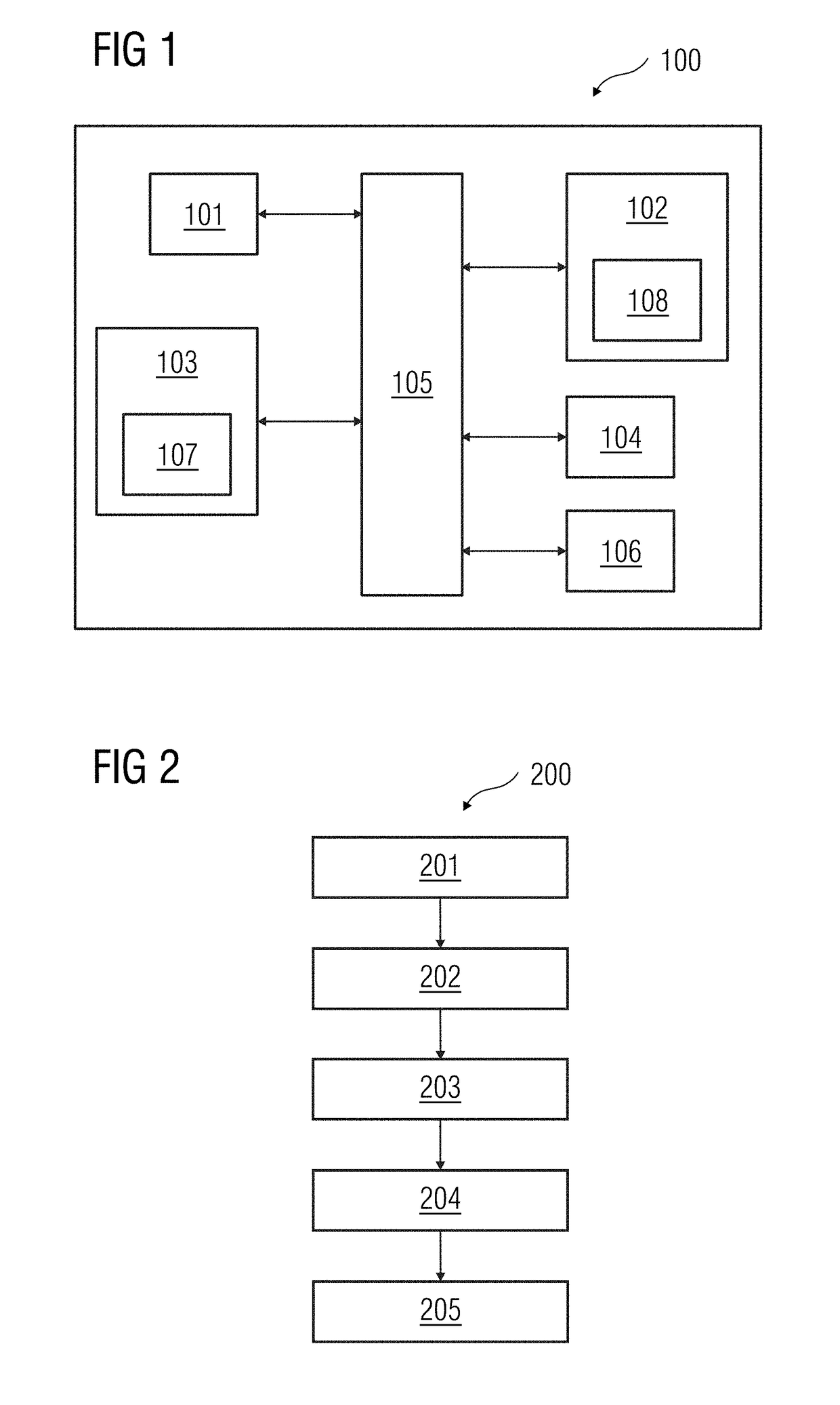
[0031]FIG. 1 is a block diagram of a data processing system 100 in which an embodiment may be implemented, for example, as a system 100 for obtaining true shape of an object, configured to perform the processes as described therein. In FIG. 1, the data processing system 100 includes a processor 101, a memory 102, a storage unit 103, an input unit 104, an output unit 106 and a bus 105.

[0032]The processor 101, as used herein, refers to any type of computational circuit, such as, but not lim...


PUM


Abstract

A method and system are provided for obtaining a true shape of objects in a medical image. The method includes receiving X-ray stand geometry parameter values, an Anglepoint, and an Anglestand values; selecting a plurality of pixels representing an object in the X-ray image; and determining magnification of each pixel in the X-ray image, that enables accurate determination of size and shape of imaged objects. The method may include determining an actual size of the object in the X-ray image and selectively reshaping the object in the X-ray image to obtain a true shape of the object.

Description

[0001]The application claims the benefit of European Patent Application No. EP 17191582.0, filed Sep. 18, 2017, which is hereby incorporated by reference in its entirety.FIELD OF TECHNOLOGY[0002]The present disclosure relates to a method and hardware and a software system of obtaining a true shape of an object in a medical image. The method includes selecting a plurality of pixels representing an object in the medical image and determining magnification of each pixel of the object in the medical image.BACKGROUND[0003]Pixel spacing represents the physical distance between two adjacent pixels. In order to measure an object in a medical image, such as an X-ray image, the pixel spacing has to be accurately determined. Image calibration techniques well known in the prior art determine the pixel spacing by assuming the patient to be positioned in the two-dimensional plane perpendicular to the central axis of the X-ray beam. Determination of pixel spacing in an X-ray image may be performed...

Claims


Application Information

Patent Timeline
Patent Type & Authority Applications(United States)
IPC IPC(8): G06T5/00G06T7/62
CPCG06T5/006G06T7/62G06T2207/10116G06T2207/10112A61B6/585
Inventor TRIPATHI, GAURAVBETHARIA, MAYUR
Owner SIEMENS HEALTHCARE GMBH
Why Eureka
Try Eureka