Enhancing pigmentation in dermoscopy images

a technology of dermoscopy and pigmentation, applied in image enhancement, color signal processing circuits, instruments, etc., can solve the problems of fogging the view, less control of illumination and image capture environment, and even greater challenges, so as to reduce or enhance the appearance of pigment components

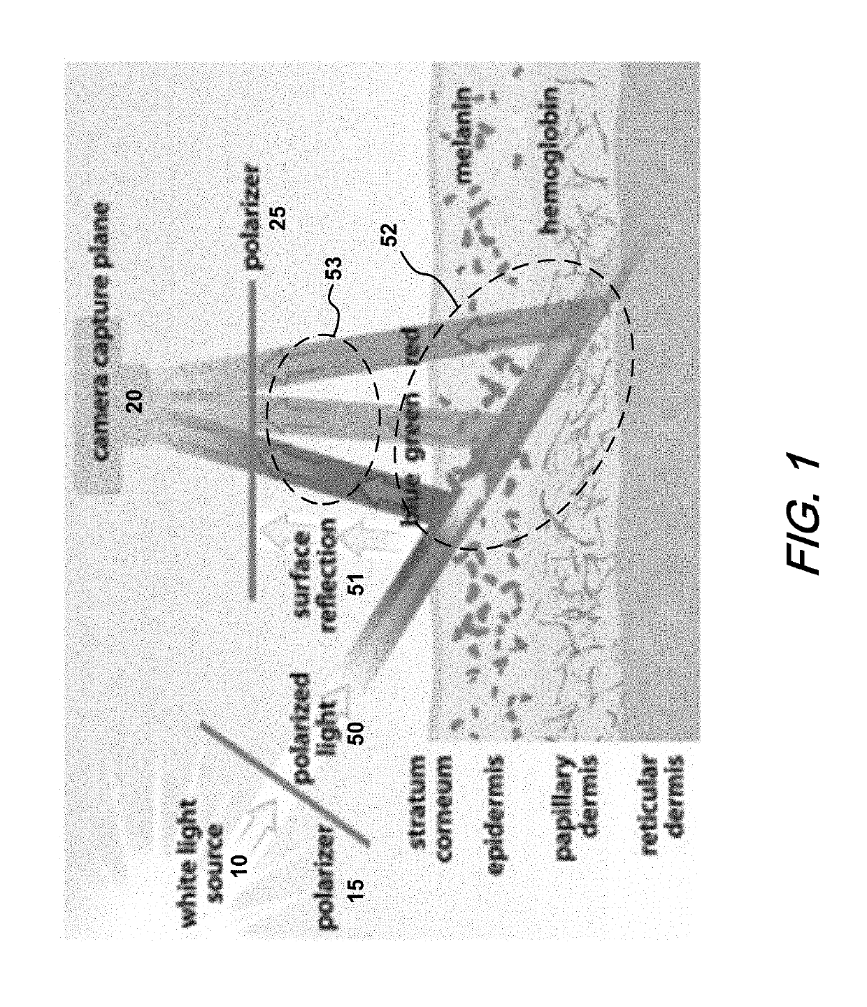
Active Publication Date: 2019-05-09
CANFIELD SCI
View PDF2 Cites 12 Cited by
  • Summary
  • Abstract
  • Description
  • Claims
  • Application Information

AI Technical Summary

Benefits of technology

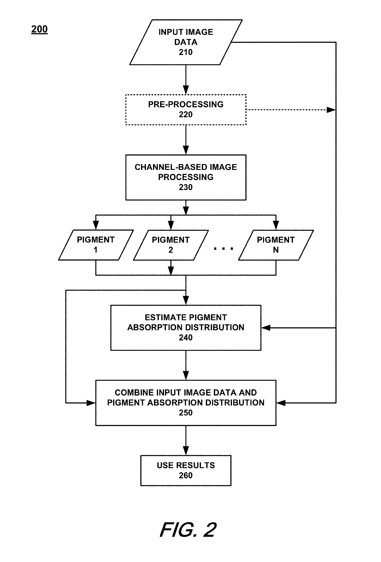
[0013]The present disclosure sets out a method performed by a skin imaging apparatus comprising: obtaining a diffuse reflectance image of an area of skin; determining a pigment component of the image; modifying the image by reducing or enhancing the appearance of the pigment component in the image; and causing the modified image to be displayed, stored, transmitted or further processed.
[0014]The present disclosure also sets out a skin imaging apparatus comprising: a storage device containing instructions; and a processor executing the instructions to: obtain a diffuse reflectance image of an area of skin; determine a pigment component of the image; modify the image by reducing or enhancing the appearance of the pigment component in the image; and cause the modified image to be displayed, stored, transmitted or further processed.

Problems solved by technology

Separating melanin structures from vascular structures, visualizing and identifying patterns in dark areas (due to concentration or depth of the pigment in these areas), and demarcating the lesion boundary, especially when the lesion is non-melanocytic, atypical, or cancerous, can be extremely challenging, even with good quality, contact cross-polarized dermoscopic imaging.
The challenge is even greater with imaging arrangements in which the illumination and image capture environment is less well-controlled, as is typical, for example, of conventional reflectance imaging.
In addition to requiring the application of fluid to the skin surface, the fluid may have bubbles which will obscure information and can fog the view, among potential drawbacks.
Due to restrictions on imaging geometry, image acquisition and processing, quality of image acquisition and illuminating optical components, polarizing filter misalignment, and / or calibration errors, it may not always be possible to capture a pure diffuse reflection image using cross-polarized imaging.
The quality of the cross-polarized data tends to degrade for shallow angles of illumination or near-perpendicular angles for detection with respect to the imaging surface, such as when capturing multiple images from various angles for 3D imaging using stereo techniques.
Cross-polarization is also compromised if the area being imaged is not flat but has appreciable curvature.
This limits the ability to obtain tissue pigmentation information using techniques such as those mentioned.
Without such systems, however, it is not typically possible to obtain diffuse reflection images of sufficient quality from which good tissue pigmentation information can be extracted.

Method used


Image

  • Enhancing pigmentation in dermoscopy images
  • Enhancing pigmentation in dermoscopy images
  • Enhancing pigmentation in dermoscopy images

Examples


Embodiment Construction

[0020]The following merely illustrates the principles of the disclosure. It will thus be appreciated that those skilled in the art will be able to devise various arrangements which, although not explicitly described or shown herein, embody the principles of the disclosure and are included within its spirit and scope. More particularly, while numerous specific details are set forth, it is understood that embodiments of the disclosure may be practiced without these specific details and in other instances, well-known circuits, structures and techniques have not been shown in order not to obscure the understanding of this disclosure.

[0021]Furthermore, all examples and conditional language recited herein are principally intended expressly to be only for pedagogical purposes to aid the reader in understanding the principles of the disclosure and the concepts contributed by the inventor(s) to furthering the art and are to be construed as being without limitation to such specifically recite...


PUM


Abstract

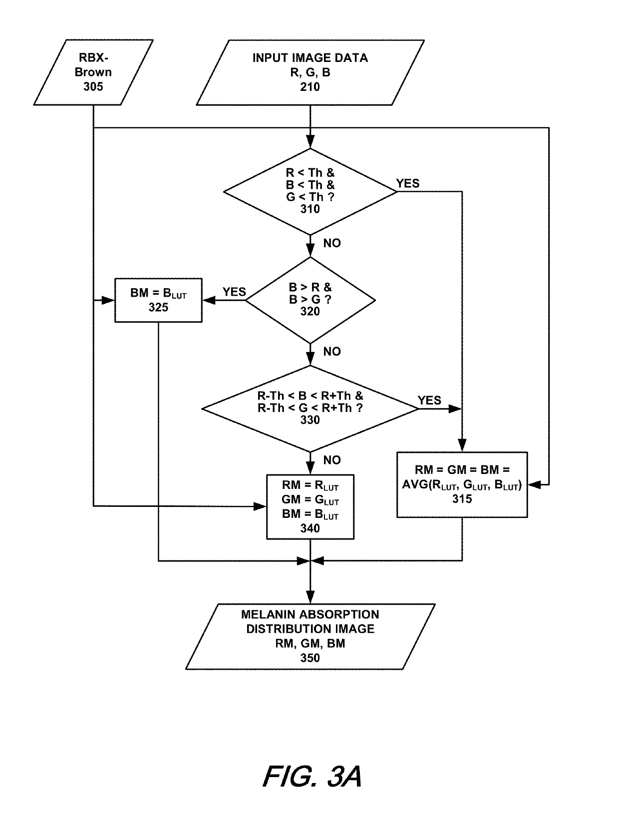
Methods and apparatuses are disclosed for modifying images of skin so as to reduce or enhance the appearance of component pigments, such as melanin and hemoglobin. A diffuse reflectance image of skin, such as a cross-polarized contact dermoscopy image, which conveys information regarding subsurface features of the skin, is processed so as to extract pigment distribution information, which is then used to correct the diffuse reflectance image, such as by reducing the appearance of melanin to allow better visualization of hemoglobin-related structures, such as vasculature. Alternatively, the diffuse reflectance image can be corrected so as to reduce the appearance of hemoglobin to allow better visualization of melanin-related structures.

Description

RELATED APPLICATIONS[0001]This Application claims priority from U.S. Provisional Patent Application No. 62 / 582,904, filed Nov. 7, 2017 and incorporated herein by reference in its entirety.BACKGROUND INFORMATION[0002]Dermoscopy is the analysis of the primary morphology of microscopic subsurface skin structures that are not visible to the unaided eye. When examining a skin lesion, a physician will typically evaluate the presence or absence of specific colors and / or structures, color and structure distribution, and whether the structure morphology is typical / regular versus atypical / irregular. A contact cross-polarized image is often used for such an evaluation.[0003]Nuances of color are important when evaluating a lesion. The colors the physician tries to identify and distinguish are Black, Brown, Gray, Blue, White, Red, Pink, and Yellow. Of these colors, Black, Brown, Gray, Blue, and White are associated with melanin distribution and its sub-surface depth. In dermoscopy images, melani...

Claims


Application Information

Patent Timeline
Patent Type & Authority Applications(United States)
IPC IPC(8): A61B5/00G06T7/90H04N5/225G06T7/00H04N9/04H04N9/64A61B5/103H04N1/60
CPCA61B5/443G06T7/90H04N5/2256G06T7/0012H04N9/04H04N9/646A61B5/0077A61B5/742A61B5/1032H04N1/6052A61B5/0013G06T2207/30088G06T2207/20092G06T2207/10024G02B27/288A61B2576/02A61B5/442G06T5/008G01N2800/207A61B5/0064A61B5/441A61B5/444H04N23/10H04N23/56
Inventor PATWARDHAN, SACHIN V.
Owner CANFIELD SCI
Why Eureka
Try Eureka