Method and apparatus for coaptive ultrasound gastrostomy

a coaptive ultrasound and gastrostomy technology, applied in the direction of balloon catheters, surgical needles, catheters, etc., can solve the problems of difficult operation, difficult to achieve, and difficult to move the conduit to the intended location

Active Publication Date: 2019-06-06
UNIV OF MARYLAND
View PDF0 Cites 1 Cited by
  • Summary
  • Abstract
  • Description
  • Claims
  • Application Information

AI Technical Summary

Benefits of technology

[0012]By allowing visual confirmation through the use of readily-available ultrasound equipment, internal catheter, conduit, or other elongate medical member placement can be achieved without the need for expensive, specialized equipment, such as endoscopes, fluoroscopes, and CT scanners. Thus, the system and method disclosed herein will enable acute care physicians, such as emergency medicine physicians and critical care physicians (i.e., intensivists), or other healthcare providers trained in the art of ultrasound, to place such catheters, conduits, and other elongate medical members safely and reliably.
[0013]With regard to certain aspects of an embodiment of the invention, the system and method may be used for inserting gastrostomy tubes in a simple bedside procedure without requiring the use of specialized consultants or specialized equipment including endoscopes, fluoroscopes, and CT scanners. The system and method make use of more widely available medical devices that require less specialized training to use, such as ultrasounds, feeding tubes, guide wires, and dilators. By allowing a wider population of medical operators to perform such procedures, the system and method disclosed herein offer safer, more immediate and more cost-effective care. For example, unlike endoscopes, ultrasound ensures that an adequate window for percutaneous gastrostomy tube insertion is present by providing visual confirmation that no bowel, organs, or blood vessels obstruct the cannulation tract between the skin surface and the stomach wall. Further, with this procedure, emergency medicine physicians may easily re-insert dislodged gastrostomy tubes and immediately discharge patients back to their residence instead of admitting them for traditional gastrostomy placement to be performed by a specialist consultant, thus saving time and decreasing consultation costs, hospital admission costs, patients' stress, and the risk of nosocomial infection. Geriatricians and rehabilitation physicians may likewise use the system and method disclosed herein in nursing home and rehabilitation facilities. This practice could dramatically reduce overall costs by eliminating patient transportation costs and thus avoiding all hospital fees.

Problems solved by technology

Procedures for placing such medical devices vary widely from application to application, but all carry the common aspect of presenting challenge to the medical staff in manipulating such medical devices within the patient's body to route them to their intended location and position them for their intended use at that location.
More particularly, often times medical procedures require manipulation of a catheter or other conduit through portions of the patient's body that are not easily accessible, and thus make maneuvering of the conduit to its intended location quite challenging.
The procedures for guiding such conduits to their intended locations in a patient's body can be difficult to perform and can risk serious injury to the patient if not performed properly.
One such procedure that presents significant challenges is the placement of gastrostomy tubes for patients requiring direct nutritional supplementation into the stomach.
This expensive equipment includes endoscopes, fluoroscopes, and computed tomography (CT) scanners, all of which require specialized training and skill to operate properly.
Even with skilled consultant physicians handling these procedures, complications can occur including tube misplacement, inadvertent injury to surrounding tissues during placement, infections, tube clogging, and tube dislodgement during use.
However, as the gastrostomy procedures require specialized skill in handling, emergency medicine physicians are often unable to perform the necessary procedures, and must instead call upon such specialist consultants, which adds to the overall expense and delay in treating the patient's issue, or risk of further complication or injury if someone lacking sufficient specialized skill attempts to address the issue.

Method used


Image

  • Method and apparatus for coaptive ultrasound gastrostomy
  • Method and apparatus for coaptive ultrasound gastrostomy
  • Method and apparatus for coaptive ultrasound gastrostomy

Examples


Embodiment Construction

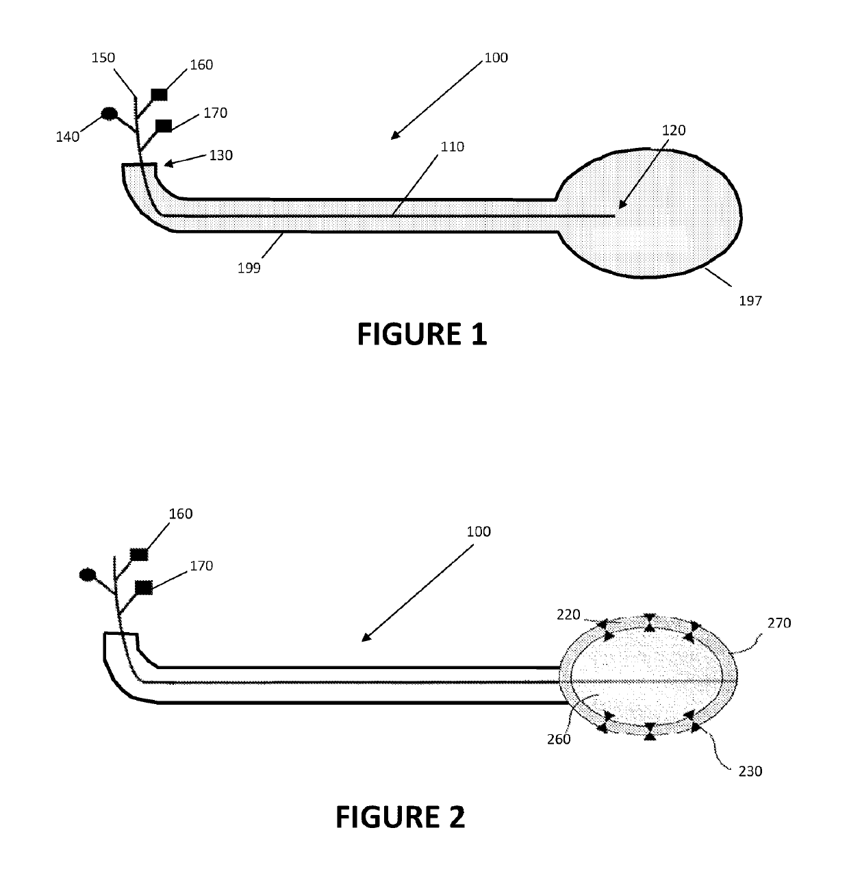
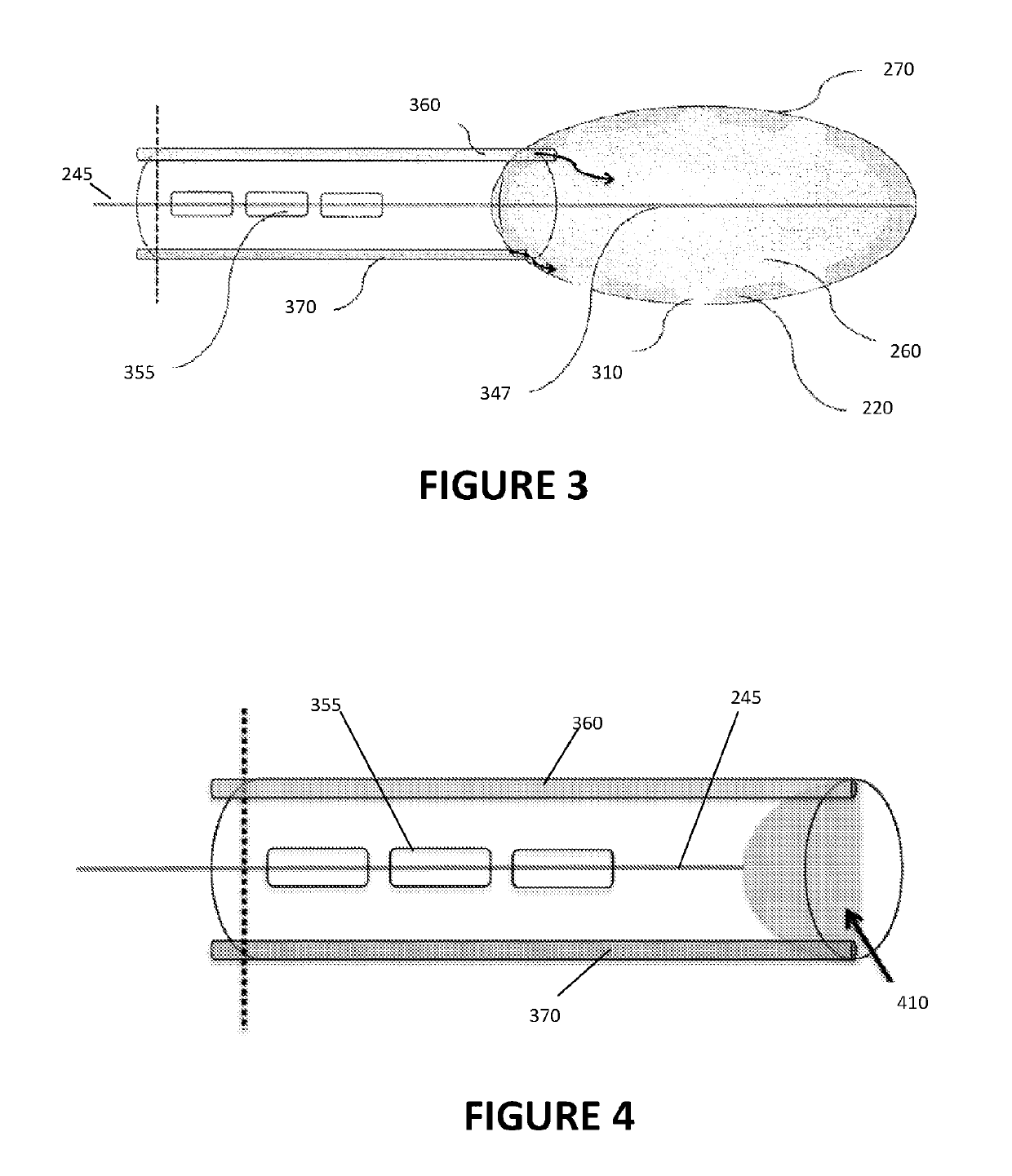
[0011]Disclosed herein is a system and method for placement of a catheter, conduit, or other elongate member, and more particularly a gastrostomy tube, within a patient's body. Such system and method are suitable for use in therapeutic interventional and / or diagnostic procedures, and may be useful for placement of medical devices, including catheters or other conduits, in varied tissue planes and cavities in a patient's body, including by way of non-limiting example the thorax, abdomen, blood vessels, and pericardium, for diagnostic, therapeutic, and / or procedural purposes. For example, such system and method may be useful in the placement of a catheter within a patient's stomach during a procedure for placement of a gastrostomy tube. Further, such system and method may be useful in the positioning of a suction tube within a patient's body to remove unwanted fluid. As used herein, all of such carriers, catheters, conduits, delivery devices, internal probes and sensors, electrodes, a...


PUM


Abstract

Disclosed is a system and method for the placement of elongate medical members within a patients body using coaptive ultrasound. In a particularly preferred embodiment, a flexible tube includes a first balloon at a distal end of the tube, and a second balloon at the distal end of the tube and positioned within the first balloon. The first and second balloons are inflatable to form one or more echogenic windows between them, which echogenic window may be detected from within a patient's body by an ultrasound probe that is external to the patient's body. Detection of such echogenic window is used to identify an acceptable location on the patient's body at which to insert a guidewire configured to receive an elongate medical member without damage to surrounding patient tissues or organs.

Description

CROSS-REFERENCE TO RELATED APPLICATIONS[0001]This application is a continuation of International PCT Application No. PCT / US2017 / 026141, entitled “Method and Apparatus for Coaptive Ultrasound Gastrostomy,” which is based upon and claims priority from co-pending U.S. Provisional Patent Application Ser. No. 62 / 318,449 entitled “Method and Apparatus for Coaptive Ultrasound Gastrostomy,” filed with the United States Patent and Trademark Office on Apr. 5, 2016, by the inventor herein, the specification of each of which is incorporated herein by reference in its entirety.FIELD OF THE INVENTION[0002]The present invention relates generally to placement of medical devices within a body in the medical field, and more particularly to methods and devices for ultrasound-guided placement of medical devices, such as catheters, conduits, carriers, electrodes, and the like, including a percutaneous gastrostomy tube, into a patient's body. Certain aspects of the invention particularly relate to method...

Claims


Application Information

Patent Timeline
Patent Type & Authority Applications(United States)
IPC IPC(8): A61B17/34A61J15/00A61M25/10
CPCA61B17/3415A61J15/0015A61M25/104A61M25/1018A61B2017/3413A61B2017/3486
Inventor TROPELLO, STEVEN
Owner UNIV OF MARYLAND
Why Eureka
Try Eureka