Apparatus and method for acquiring near infrared-based diagnostic images of teeth

a near infrared and diagnostic image technology, applied in the field of apparatus and a method of acquiring an image for dental diagnosis, can solve the problems of poor contrast in the x-ray image, difficult to detect cavities with a small volume from general x-ray images, and dangerous x-rays, so as to eliminate the risk of radiation ionization, easy to find occlusal cavities on teeth, and convenient to provide the safety and reliability of the apparatus to the patient.

Active Publication Date: 2019-12-05
KOREA PHOTONICS TECH INST
View PDF0 Cites 7 Cited by
  • Summary
  • Abstract
  • Description
  • Claims
  • Application Information

AI Technical Summary

Benefits of technology

[0020]According to the present invention, near-infrared radiation is used rather than X-rays, so that the risk of ionization caused by radiation can be eliminated, and safety and reliability for the apparatus can be provided to the patient.
[0021]Also, according to the present invention, since near-infrared radiation is used, it is easy to find occlusal cavities on teeth, or the like.
[0022]Also, according to the present invention, a panoramic image is implemented using images acquired at different angles, so that the degree of twisting of teeth / periodontal axes, the degree of occlusion, or the like are quantified for dental diagnosis.
[0023]Also, according to the present invention, by applying the mouthguard-shaped dental diagnosis module provided with the pressure sensor, the bite pressure when the mouthguard-shaped dental diagnosis module is bitten down on can be measured and can be imaged, whereby the bite pressure distribution for each tooth can be obtained and the hardness for each tooth, and the like are quantified for dental diagnosis.
[0024]Also, according to the present invention, a near-infrared light source, a pressure sensor, and a general image camera are used, so that the production cost and the sales price of the apparatus for acquiring a dental image can be lowered.

Problems solved by technology

X-rays are dangerous because X-rays ionize molecules of living tissue.
However, the cavities with a small volume having higher permeability or lower attenuation make only a small partial change in the intensity of the transmitted radiation, so that for the portion of the small cavities, contrast in the X-ray image is poor.
Therefore, it is difficult to detect cavities with a small volume from general X-ray images.
On the other hand, in general, there is a problem in that it is difficult to find occlusal cavities on the wide and flat chewing surfaces.
However, the X-ray method has a problem that the reliability for detecting cracks is poor.

Method used


Image

  • Apparatus and method for acquiring near infrared-based diagnostic images of teeth
  • Apparatus and method for acquiring near infrared-based diagnostic images of teeth
  • Apparatus and method for acquiring near infrared-based diagnostic images of teeth

Examples


Embodiment Construction

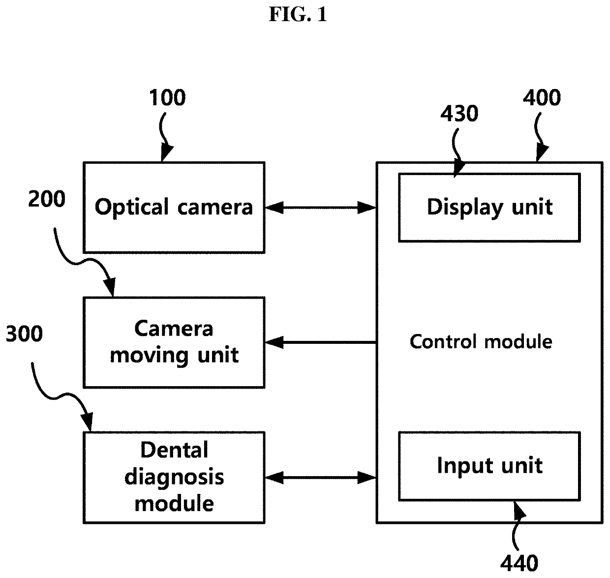
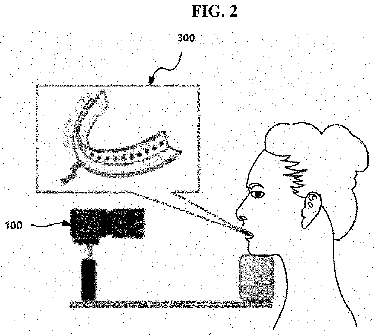
[0034]Hereinafter, a configuration and an operation of an apparatus for acquiring a near-infrared image for dental diagnosis according to the present invention will be described with reference to the accompanying drawings, and a method of acquiring the image for dental diagnosis, which is performed by the apparatus, will be described.

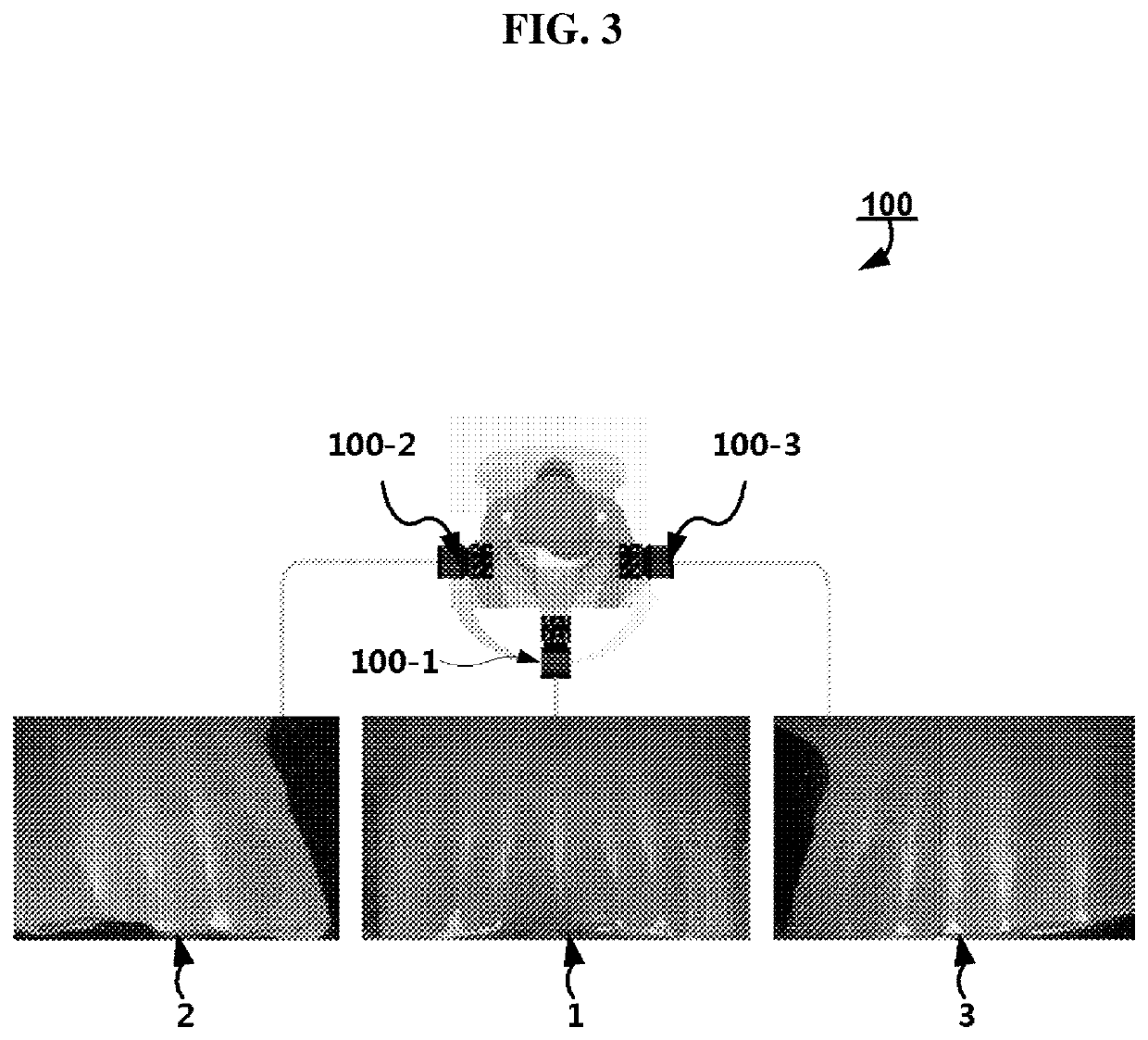
[0035]FIG. 1 is a diagram illustrating a configuration of an apparatus for acquiring a near-infrared image for dental diagnosis according to the present invention. FIG. 2 is a diagram illustrating an application example of an apparatus for acquiring a near-infrared image for dental diagnosis according to the present invention. FIG. 3 is a diagram illustrating images acquired by performing photographing from the front, left, and right with an apparatus for acquiring a near-infrared image for dental diagnosis according to an embodiment of the present invention. FIG. 4 is a diagram illustrating a physical configuration of a mouthguard-shaped dental diagnos...


PUM


Abstract

The present invention relates to an apparatus and a method of acquiring an image for dental diagnosis. More particularly, the present invention relates to an apparatus and a method of acquiring a near-infrared image for dental diagnosis, wherein an image for dental diagnosis is acquired using near-infrared radiation and a general image camera rather than using X-rays.

Description

CROSS-REFERENCE TO RELATED APPLICATIONS[0001]The present application is a continuation of International Patent Application No. PCT / KR2018 / 000771, filed Jan. 17, 2018, which is based upon and claims the benefit of priority to Korean Patent Application No. 10-2017-0074574, filed on Jun. 14, 2017. The disclosures of the above-listed applications are hereby incorporated by reference herein in their entirety.TECHNICAL FIELD[0002]The present invention relates to an apparatus and a method of acquiring an image for dental diagnosis. More particularly, the present invention relates to an apparatus and a method of acquiring a near-infrared image for dental diagnosis, wherein an image for dental diagnosis is acquired using near-infrared radiation and a general image camera rather than using X-rays.BACKGROUND ART[0003]Generally, in dentistry, before a dental treatment, a mouth portion of a patient is subjected to radiography such that an X-ray image showing patient's teeth structure and conditi...

Claims


Application Information

Patent Timeline
Patent Type & Authority Applications(United States)
IPC IPC(8): A61B5/00H04N5/232H04N5/225G06T7/00A61B1/24A61B5/11
CPCH04N5/2256G06T7/0012A61B5/1111H04N5/23238G06T2207/30036A61B1/24G06T2207/10048A61B2562/0247A61B5/0088H04N5/23299H04N5/33A61B1/0661A61B1/04A61B1/045A61B1/00009A61B1/00045A61B5/038A61B1/046H04N23/56G06T5/50H04N23/695H04N23/698
Inventor LEE, BYEONG-ILSHIN, IN-HEE
Owner KOREA PHOTONICS TECH INST
Why Eureka
Try Eureka