
Osteotomy Device

Inactive Publication Date: 2020-04-09
WALZMAN DANIEL EZRA
View PDF18 Cites 1 Cited by
  • Summary
  • Abstract
  • Description
  • Claims
  • Application Information

AI Technical Summary

Benefits of technology

The present invention is a device that combines multiple osteotomy tools to create a single device that can perform all the necessary functions of cutting, irrigation, aspirating, and cauterizing while providing live video information to the surgeon. The device is designed to minimize tissue injury and can be adjusted and directed to the targeted surgical site.

Problems solved by technology

While Image guided surgery typically employs electromagnetic sensors / tracking systems, radiofrequency electromagnetic sensors (e.g., electromagnetic coils) which are placed on the surgical instruments and on a localizer frame worn by the patient, they do not have built in irrigation and respiration systems.
More particularly, this shortcoming results in the requirement that the surgeon insert and remove separate irrigation and respiration systems causing delay in procedures.
The presence of said separate systems causes sensors which have been mounted on proximal portions of the instruments (e.g., on the handpiece of the instrument) to be moved away from targeted surgery sites resulting in the reduction of the accuracy of sensor information.
Currently only select balloon sinu-plasty procedures are performed in this setting, but these are less effective at relieving symptoms, and have much higher rates of recurrent symptomatic sinus blockage / infections.
While some use of insertable shielding is described in the prior art, such shielding is difficult to position, tends to migrate out of position, and can cause irritation to tissue.

Method used


Image

  • Osteotomy Device
  • Osteotomy Device
  • Osteotomy Device

Examples


Embodiment Construction

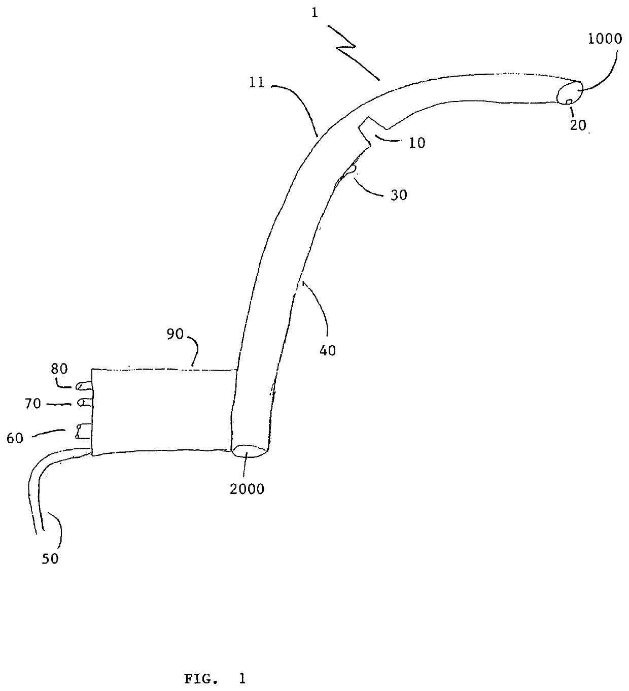
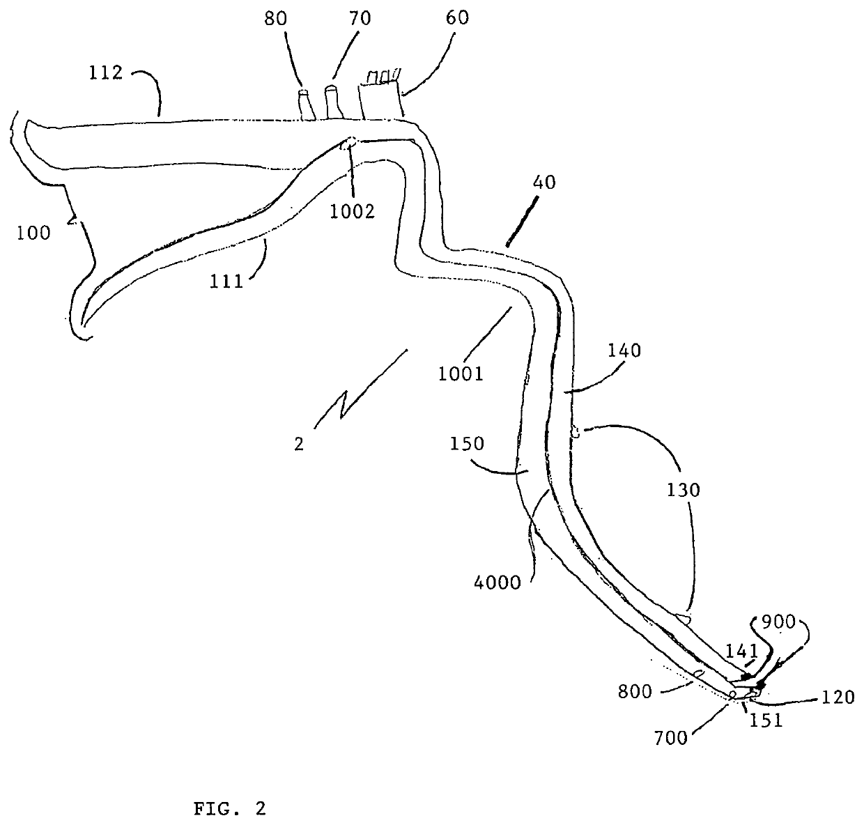
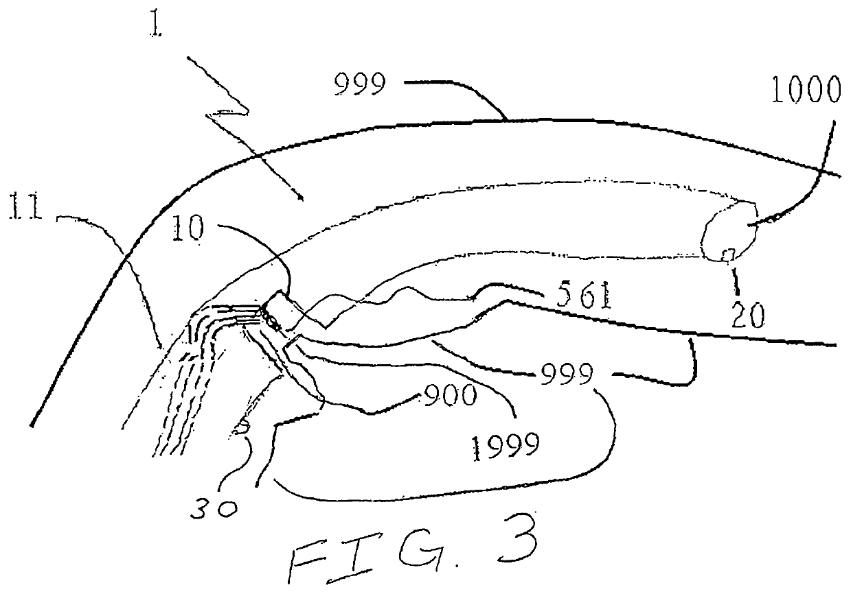
[0014]Now referring to FIG. 1, herein is described an osteotomy device 1 is composed of an elongate, straw-like cylinder 11 with proximal opening 2000 located outside the patient's body, and distal hole 1000 located inside the patient's body. Between proximal hole 2000 and distal hole 1000 is a side-slit 10 along the outside of cylinder element 11. Additionally, device 1 includes a handle 90, attached to cylinder 11. Handle 90 is optionally detachable when element 2 (illustrated in FIG. 2) is inserted through cylinder 11 via proximal hole 2000, and thereby rendering optional handle 90 redundant.

[0015]More particularly, cylinder 11 is composed of hard but flexible material suitable for protecting tissue proximal to a target surgical site. It is formed, optionally, of a biocompatible metal, plastic, or other suitable material, that is preferentially but optionally malleable. The cylinder including at least one side slit 10, wherein side slit 10 serves as a conduit to facilitate an ost...


PUM


Abstract

The present disclosure teaches a novel medical device for osteotomy, capable of simultaneous aspiration, cauterization, image-acquisition, irrigation and tissue-protection, adapted for ear, nose and throat procedures, but available for other cavity surgeries.

Description

FIELD OF THE INVENTION[0001]The described invention relates generally to medical devices, systems and methods used for surgery to cut, excise or otherwise shape bones, and more particularly to methods and devices for performing image guided interventional procedures to treat disorders of the paranasal sinuses, ears, nose or throat (ENT).BACKGROUND OF THE INVENTIONPrior Art[0002]Osteotomy devices are numerous. They include devices for cauterizing, irrigating, aspirating, image-collection, and cutting, among others. These devices are generally standalone, and normally can only be used in pairs, occasionally threes. The imaging is of two types: the first involves external sensors with tagged instruments; the second involves image-collection, either prior to the surgery or mounted on an insertion device.[0003]Image-guided surgical techniques and devices were developed for neurosurgery and have now been adapted for use in certain ear, nose and throat surgeries, including sinus surgeries....

Claims


Application Information

Patent Timeline
IPC IPC(8): A61B90/00A61B1/00A61B1/015A61B1/05A61B17/15
CPCA61B1/00071A61B90/04A61B1/00009A61B1/05A61B17/151A61B2018/00595A61B1/00147A61B90/37A61B1/015A61B1/00119A61B2217/005A61B2217/007A61B17/24A61B2090/371A61B2017/00946A61B90/30A61B17/320068A61B17/3421A61B90/361A61B2017/003A61B2017/3445A61B1/00181A61B1/233A61B1/00087
Inventor WALZMAN, DANIEL EZRA
Owner WALZMAN DANIEL EZRA
Learn More