System and method for assessing breast cancer risk using imagery

a breast cancer risk and imaging technology, applied in image enhancement, healthcare informatics, instruments, etc., can solve the problems of breast cancer risk increase, breast cancer risk later diagnosis, and increased so as to achieve high accuracy, reduce the risk of breast cancer, and add substantial cost or effort.

Active Publication Date: 2020-05-28
ICAD INC
View PDF0 Cites 8 Cited by
  • Summary
  • Abstract
  • Description
  • Claims
  • Application Information

AI Technical Summary

Benefits of technology

[0007]This invention overcomes disadvantages of the prior art by providing an easily implementable prediction tool for individualized breast cancer screening without adding substantial cost or effort to the health care system, that can be based on detection of structures in the tissue, such as microcalcifications. Such microcalcifications can be based (e.g.) on detection of at-risk lesions. The characteristics of these detected structures can be optionally combined with risk factors, including, but not limited to, with mammographic density, genetic risk variants, inherited risks, comorbidities, hormonal and lifestyle factors. The process(or) employed includes a CAD (or similarly configured and functioning) computing system, based upon a deep learning, AI, neural network, and / or similar computing / data-handling arrangement, which can produce highly accurate features from acquired imagery. Notably, the CAD system for at-risk lesions is employed to augment and facilitate a prediction of risk of developing a disease (e.g. breast cancer) in addition to more traditional applications, in which the CAD is employed to diagnose existing conditions. More particularly, the acquired breast image data can provide additional image features that are, in turn, used to derive more informative predictive risk score models than previously employed microcalcifications and density (masses) measurements.

Problems solved by technology

Women with dense breasts have both an increased risk of breast cancer and a higher probability for a cancer being masked (undetected).
Women with abnormal tissue changes in the breast have increased risk for later diagnosis of breast cancer.
These models do not take breast imagery for at-risk lesions into consideration.

Method used


Image

  • System and method for assessing breast cancer risk using imagery
  • System and method for assessing breast cancer risk using imagery
  • System and method for assessing breast cancer risk using imagery

Examples


Embodiment Construction

I. System Overview

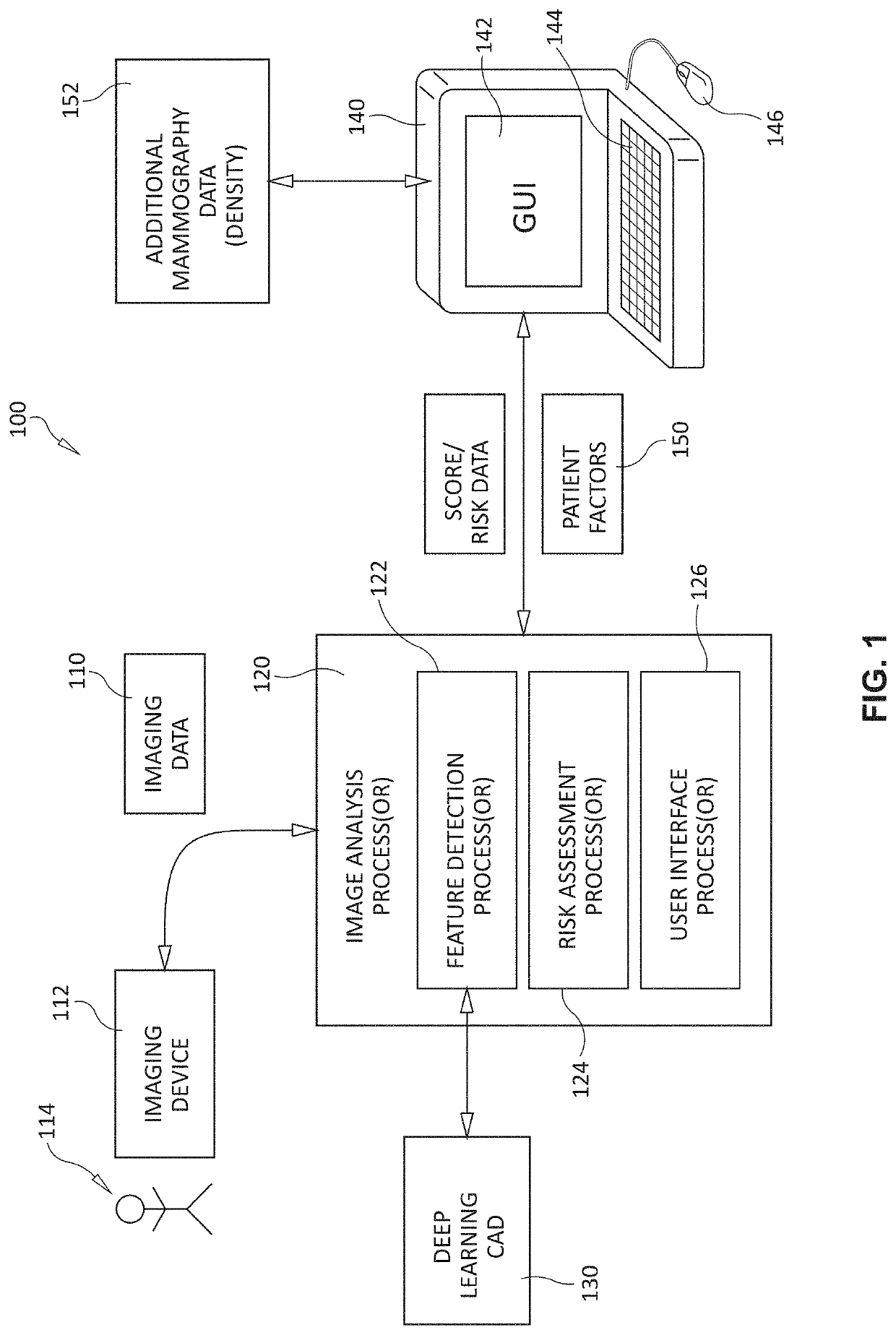
[0021]Reference is made to FIG. 1, which shows a generalized arrangement 100 in which image data 110 is derived from an imaging device 112 or store of acquired imaged—for example those acquired from a scan of a patient 114 based upon (e.g.) mammography. The image data 110 is provided to a processor and associated analysis process 120. The process(or) 120 is part of a deep learning computer-aided detection (CAD) system 130 that builds and / or employs neural networks, or similar learning arrangements / data structures (e.g. AI-based systems) to derive scores for use in evaluating the significance of detected structures within the image and underlying tissue. Note, that as used herein, the term “CAD” can refer to a computing system (hardware, software and / or firmware) that performs computer-aided detection processes, and can also (optionally) perform computer-aided analysis processes in a manner clear to those of skill. In addition, the term “CAD” can be taken to include...


PUM


Abstract

This invention provides a system and method for assessing risk of a breast cancer diagnosis based upon imagery of tissue and (optionally) other patient-related factors. A CAD (or similar) system analyzes the imagery and generates a plurality of numerical feature values. An assessment module receives inputs from patient factors and history and computes the risk based upon the feature values and the patient factors and history. A masking module receives inputs from the patient factors and history, and computes the risk of having a cancer, which cancer is otherwise characterized by a low probability of detection, based upon the feature values and the patient factors and history. A recall module receives inputs from the assessment module and the masking assessment module, and generates a computer-aided indication of a clinical follow-up by the patient. Results of assessment(s) can be displayed to the clinician and/or patient using a graphical interface display.

Description

RELATED APPLICATION[0001]This application claims the benefit of U.S. Provisional Application Ser. No. 62 / 770,958, entitled SYSTEM AND METHOD FOR ASSESSING BREAST CANCER RISK USING IMAGERY, filed Nov. 23, 2018, the teachings of which are incorporated herein by reference.FIELD OF THE INVENTION[0002]This invention relates to systems and methods to determine risk for being diagnosed with a cancerous condition in (e.g.) the breast, based on imagery of breast tissue and other tissues, and more particularly to systems and methods that can also incorporate genetic risk variants, inherited risks, personal history, and / or lifestyle factors.BACKGROUND OF THE INVENTION[0003]Risk prediction models for breast cancer use lifestyle factors, family history of breast cancer, mammographic density, genetic determinants, or any combination of these factors to predict risk of developing the disease. Mammographic density is one of the strongest risk factors for breast cancer and consists of the radiograph...

Claims


Application Information

Patent Timeline
Patent Type & Authority Applications(United States)
IPC IPC(8): G06T7/00G16H30/20G16H50/30
CPCG16H50/30G06T2207/30068G06T2200/24G06T2207/30096G16H30/20G06T2207/10116G06T7/0012G16H50/20G16H20/10G16H30/40G06Q50/163G06Q10/063
Inventor HALL, PER FRANS LEONARDERIKSSON, MIKAEL EMIL DANJAFARI-KHOUZANI, KOUROSHPERIASWAMY, SENTHIL
Owner ICAD INC
Why Eureka
Try Eureka