
Intraoral scanner

a scanner and intraoral technology, applied in the field of dental scanners, can solve the problems of large form factor and bulkiness

Active Publication Date: 2020-07-02
DENTLYTEC G P L
View PDF7 Cites 4 Cited by
  • Summary
  • Abstract
  • Description
  • Claims
  • Application Information

AI Technical Summary

Benefits of technology

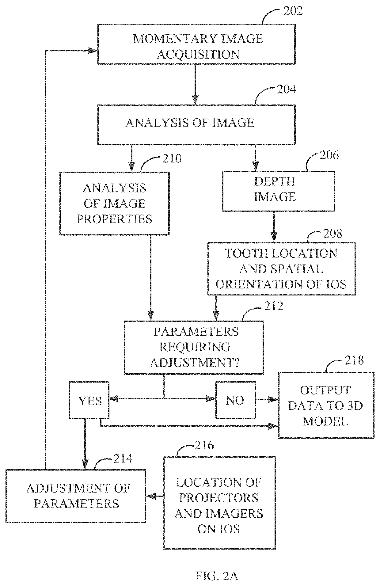
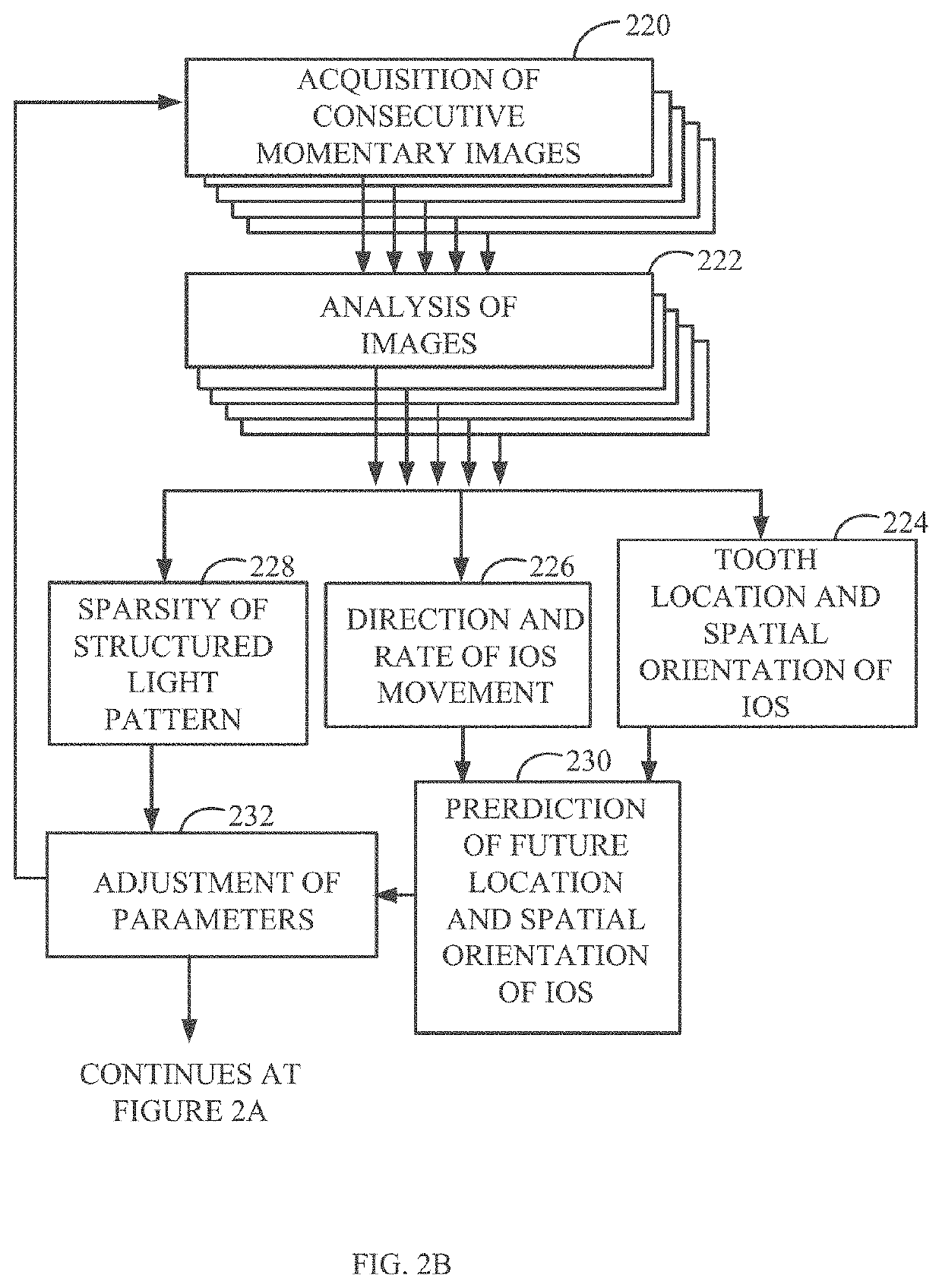
The patent describes a method for improving image quality by adjusting image acquisition parameters based on the position of a specific location on an object. This allows for more accurate positioning of the location in the image.

Problems solved by technology

However, commonly used IOSs combine the use of image sensors, light emitters, several lenses, mirrors, reflectors, beam splitters and other components the size and quantity of which contribute to a relatively large form factor and bulkiness of the IOS.

Method used


Image

  • Intraoral scanner
  • Intraoral scanner
  • Intraoral scanner

Examples


Embodiment Construction

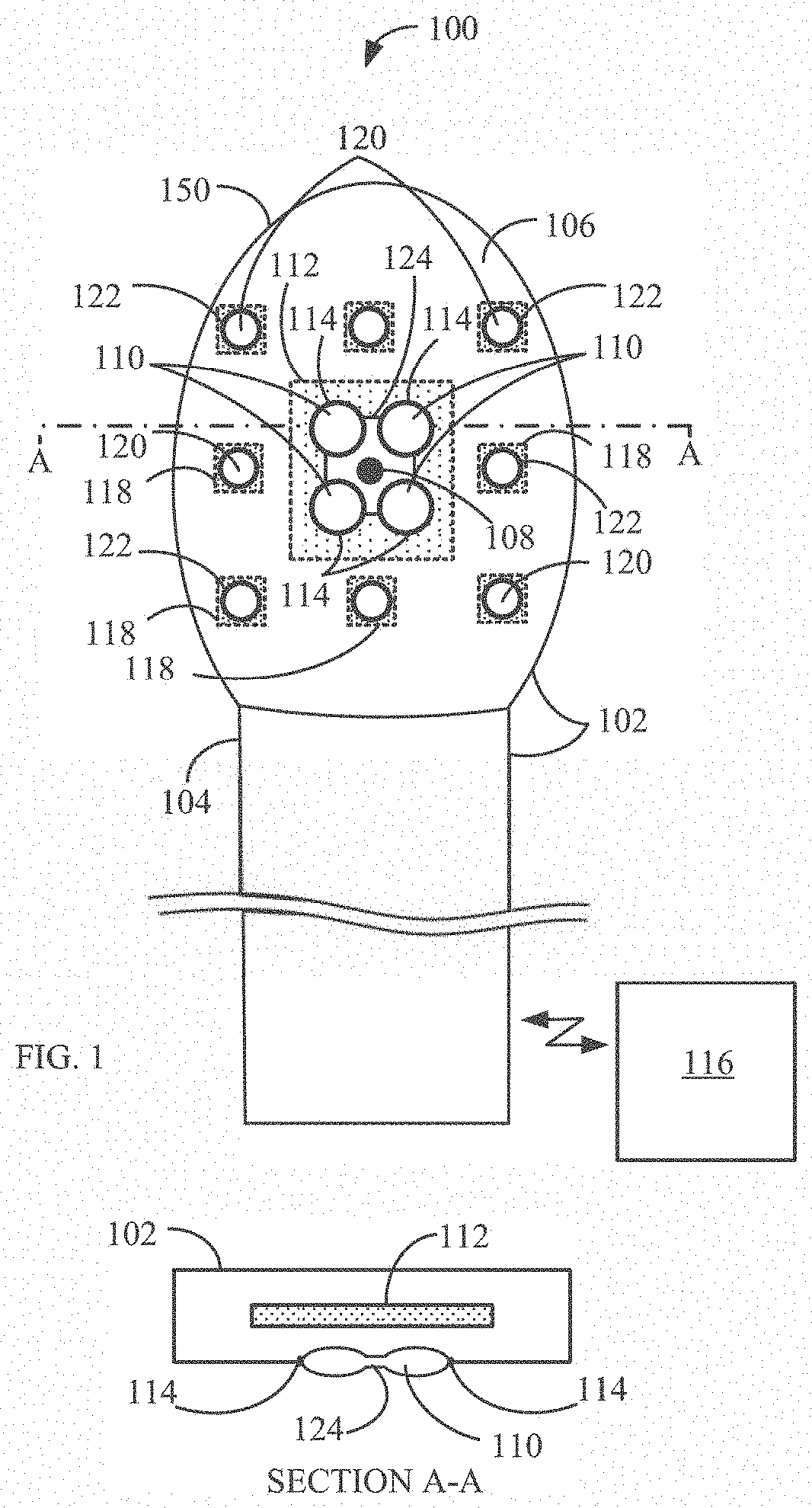
[0007]According to an aspect of some embodiments of the disclosure, there is provided a method of scanning an oral cavity comprising: acquiring, using an intraoral scanner (IOS) head, without changing a position of said IOS head, a first image of a first region of interest (ROI) and a second image of a second ROI where said first and said second ROIs are of different portions of a dental arch of the oral cavity and do not overlap; reconstructing depth information for said first and said second ROI; and generating a single model of said dental arch by combing said depth information.

[0008]According to some embodiments of the disclosure, the acquiring of said first image and said second image is simultaneous.

[0009]According to some embodiments of the disclosure, the first ROI is captured by a first IOS field of view (FOV) and said second ROI is captured by a second IOS FOV.

[0010]According to some embodiments of the disclosure, the IOS includes at least two imagers where said first FOV ...


PUM


Abstract

A method of scanning an oral cavity including: acquiring, using an intraoral scanner (IOS) head, without changing a position of the IOS head, a first image of a first region of interest (ROI) and a second image of a second ROI where the first and the second ROIs are of different portions of a dental arch of the oral cavity and do not overlap; reconstructing depth information for the first and the second ROI; and generating a single model of the dental arch by combing the depth information.

Description

[0001]This application claims the benefit of priority under 35 USC § 119(e) of U.S. Provisional Patent Application No. 62 / 537,340 filed 26 Jul. 2017, the contents of which is incorporated herein by reference in its entirety.[0002]This application is also related to PCT Patent Application No. PCT / IL2016 / 050058, titled “SYSTEM, DEVICE, AND METHOD FOR DENTAL INTRAORAL SCANNING” filed on Jan. 18, 2016, which claims the benefit of priority under 35 USC § 119(e) of U.S. Provisional Patent Application No. 62 / 104,835, filed on Jan. 18, 2015.[0003]This application is also related to: Provisional Patent Application No. 62 / 364,681, filed on Jul. 4, 2017; Provisional Patent Application No. 62 / 528,496, filed on Jul. 20, 2016; U.S. Pat. No. 9,204,804; U.S. Patent publication No. 2010-0189341; U.S. Pat. No. 8,971,999; and “Application of Intra-Oral Dental Scanners in the Digital Workflow of Implantology”, by Wicher J. van der Meer1*, Frank S. Andriessen2, Daniel Wismeijer3, Yijin Ren4.FIELD AND BA...

Claims


Application Information

Patent Timeline
Patent Type & Authority Applications(United States)
IPC IPC(8): A61C9/00A61C13/00A61C7/00G06K9/00G06K9/20G06T19/20
CPCG06T19/20A61C9/006G06T2219/2004A61C7/002A61C9/0066G06K9/00214A61C13/0004G06K9/2036G06K9/2054A61B5/0088A61B5/4547A61B2560/0223A61B2562/0238A61B2576/02G01B11/24G01B11/2518G01B11/2545G01B2210/52G16H30/40G06T7/521G06T7/579G06T7/593G06T2207/10016G06T2207/10048G06T2207/30036G06T2200/08G06V20/653
Inventor PESACH, BENNYREUVENNY, AMITAI
Owner DENTLYTEC G P L
Learn More