
Use of an integrated microfluidic chip for analysis of cell motility and prediction and prognosis of patient survival

Pending Publication Date: 2020-08-06
THE JOHN HOPKINS UNIV SCHOOL OF MEDICINE
View PDF0 Cites 6 Cited by
  • Summary
  • Abstract
  • Description
  • Claims
  • Application Information

AI Technical Summary

Benefits of technology

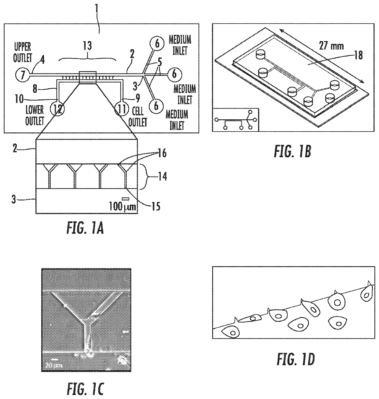
The present invention provides a method for identifying the invasiveness of brain cancer cells or predicting the outcome of a brain tumor patient by analyzing the characteristics of cancer cells obtained from the tumor. This is accomplished by using an integrated microfluidic apparatus that can incubate and image cancer cells, allowing the identification of invasive cells and the prediction of recurrence or low survival. The invention also provides a method for identifying effective chemotherapy treatments for brain cancer by testing multiple agents and monitoring their impact on cancer cell invasion. Overall, the invention provides a valuable tool for advancing brain cancer research and treatment.

Problems solved by technology

Personalized medical plans aiming to limit metastasis are difficult to develop.
However, there still is an unmet need in the ability of clinicians and investigators to identify patients with brain cancer that is highly invasive and which correlates with lower survival times and high risk of recurrence.

Method used


Image

  • Use of an integrated microfluidic chip for analysis of cell motility and prediction and prognosis of patient survival
  • Use of an integrated microfluidic chip for analysis of cell motility and prediction and prognosis of patient survival
  • Use of an integrated microfluidic chip for analysis of cell motility and prediction and prognosis of patient survival

Examples


example 1

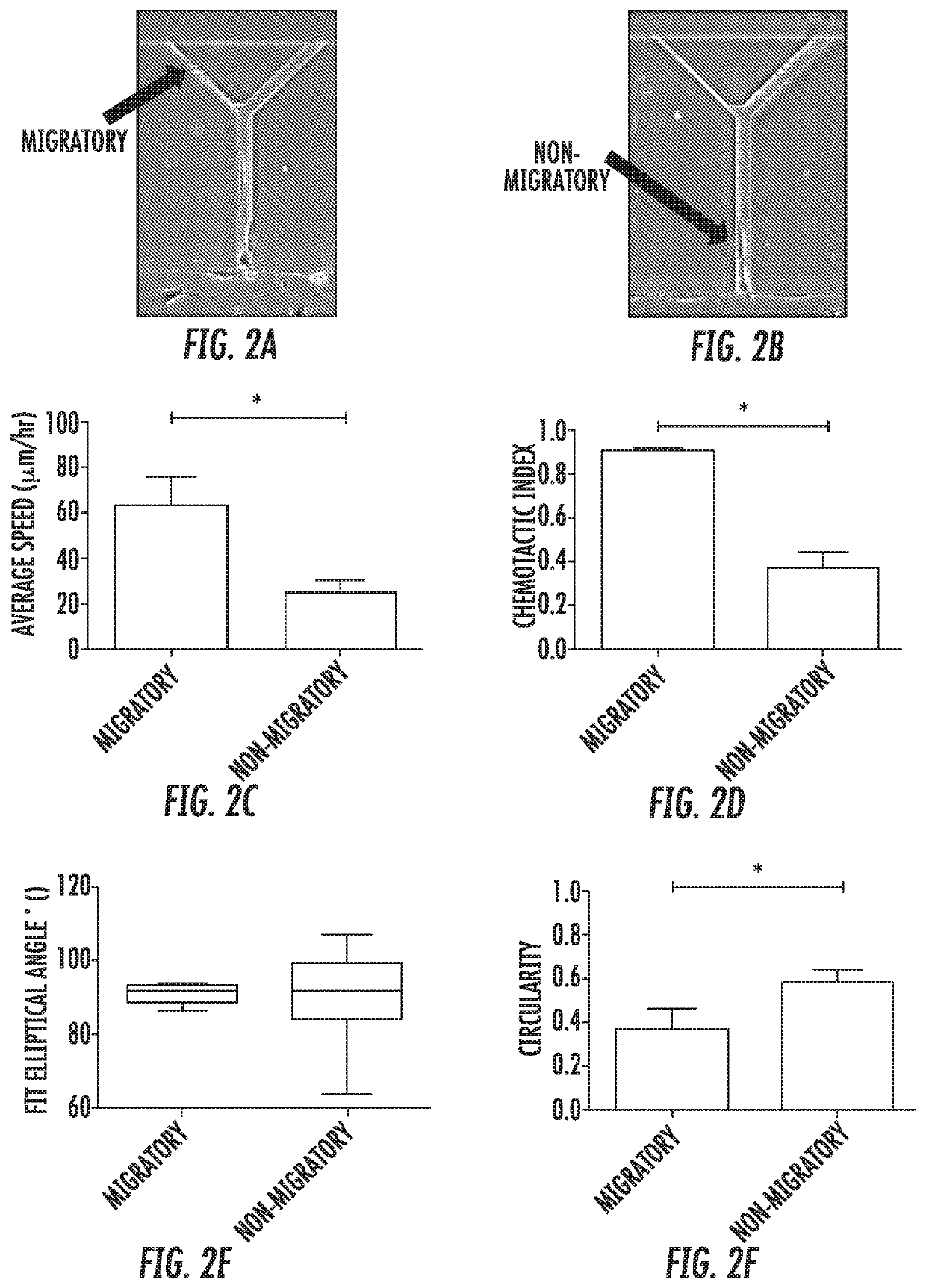
[0088]Bifurcating Channels Allow Identification of Migratory Cells: Cell tracking of all MDA-MB-231 cells within the channels revealed two distinct subpopulations: migratory and non-migratory cells (FIGS. 2A,B). 22±3% of human metastatic MDA-MB-231 breast cancer cells were migratory. Interestingly, this subpopulation correlates with the % of MDA-MB-231 (28%) bearing the CD44+ / CD24− molecular signatures that is used to define breast cancer stem cells. Migratory cells, defined as those cells reaching the branch channels, migrated more than twice as fast as non-migratory cells (FIG. 2C). Migratory cells were also significantly more directional. The chemotactic index of migratory cells increased to 0.91 in comparison to a chemotactic index of 0.37 for nonmigratory cells.

[0089]Analysis of cell shape indicated that migratory cells were aligned with and elongated along the channel wall. The fit elliptical angle of a cell perfectly aligned along the wall was 90°. Although both migratory and...

example 2

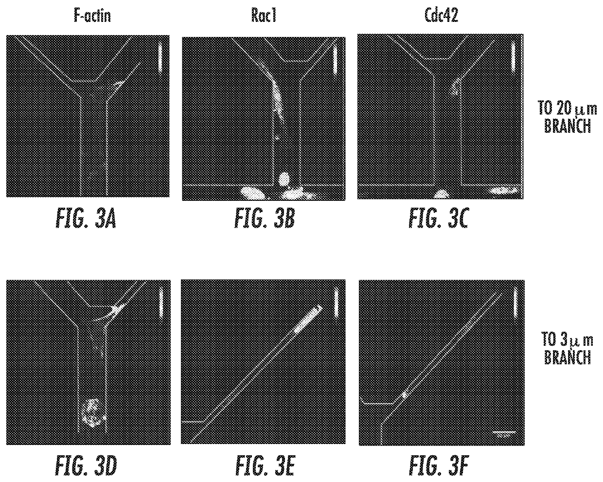
[0090]Contact Guidance Overcomes Steric Hindrance for Migratory Cells: Of those cells that were migratory, the vast majority moved preferentially along one wall of the feeder channels and remaining polarized, with a significantly lower number of changes in direction compared to non-migratory cells. Representative cell tracks illustrating this trend are shown in FIG. 4A. Interestingly, migration direction at the bifurcation was not dependent on the width of the resultant branch, even though entering the 3 μm-wide branch required significant deformation of the cell body. Instead, cells continued to be polarized and moved readily into the “branch” channel, regardless of the branch channel width (FIG. 4B). Thus, contact guidance dominated steric hindrance at these channel widths for migratory cells and was likely the driver of directed migration for this subpopulation.

example 3

[0091]PI3K Inhibition Promotes Spontaneous Migration of MDA-MB-231 Cells: There is evidence that PI3K signaling is required to stabilize nascent protrusions. New protrusions away from the wall along which a cell is migrating would discourage contact guidance. Therefore, it was investigated whether inhibiting PI3K could promote contact guidance in 200 μm-long microchannels.

[0092]Inhibition of PI3K signaling using the PI3K inhibitor LY294002 increased the migratory cell population in 200 μm-long microchannels from 25% to 65% and the ratio of contact guided cells from 66% to 93% (FIG. 4A,B). PI3K inhibition did not impact overall cell speed, as control and LY294002-treated cells moved at the same average speed (FIG. 5C). However, cells in which PI3K signaling was inhibited moved with greater directionality, as indicated by the higher chemotactic index for these cells vs. control cells (FIG. 5D). This result is consistent with the expected inhibition of nascent protrusions upon LY294002...


PUM


Abstract

The present invention describes an integrated apparatus that enables identification of invasive tumor cells directly from a specimen. The methods using the apparatus can be used to prognose or predict the survivability of the cancer in a subject and the risk of recurrence of the cancer in the subject after treatment. The methods disclosed herein can be used to determine which chemotherapeutic or other therapies most strongly inhibit the tumor cells invasiveness as a form of personalized therapy.

Description

REFERENCE TO RELATED APPLICATIONS[0001]This application claims the benefit of U.S. Provisional Patent Application No. 62 / 262,158, filed on Dec. 2, 2015, which is hereby incorporated by reference for all purposes as if fully set forth herein.STATEMENT OF GOVERNMENTAL INTEREST[0002]This invention was made with government support under grant no. NCI-U54-CA143868 awarded by the National Institutes of Health. The government has certain rights in the invention.BACKGROUND OF THE INVENTION[0003]Personalized medical plans aiming to limit metastasis are difficult to develop. The current state of the art requires the expansion of human cancers in immunodeficient mice before the cancers can be subjected to drug screenings. It is known that metastatic subpopulations of cancer cells have heightened motility which is linked to aggressiveness and invasiveness of the cancer. The ability to identify such a subpopulation of cells in a tumor of a patient would be useful in classifying the aggressivenes...

Claims


Application Information

Patent Timeline
IPC IPC(8): G01N33/574B01L3/00G01N33/50
CPCG01N33/5011B01L3/502761G01N33/5029B01L2300/0867B01L2200/0652B01L2300/0816G01N33/574G01N33/57407A61P35/00G01N33/5091G01N2800/7028
Inventor KONSTANTOPOULOS, KONSTANTINOSPAUL, COLINQUINONES-HINOJOSA, ALFREDOSHAH, SAGAR RAMESHRUIZ-VALLS, ALEJANDROYANKASKAS, CHRISTOPHERMARTINEZ-GUTIERREZ, JUAN CARLOSWONG, BIN SHENG
Owner THE JOHN HOPKINS UNIV SCHOOL OF MEDICINE
Learn More