
Systems and methods for image classification

a technology of image classification and system, applied in image analysis, image enhancement, instruments, etc., can solve the problems of inability to localize annotations, inability to obtain localized annotations, and inability to provide localized annotations

Pending Publication Date: 2020-08-06
OWKIN INC +1
View PDF0 Cites 22 Cited by
  • Summary
  • Abstract
  • Description
  • Claims
  • Application Information

AI Technical Summary

Benefits of technology

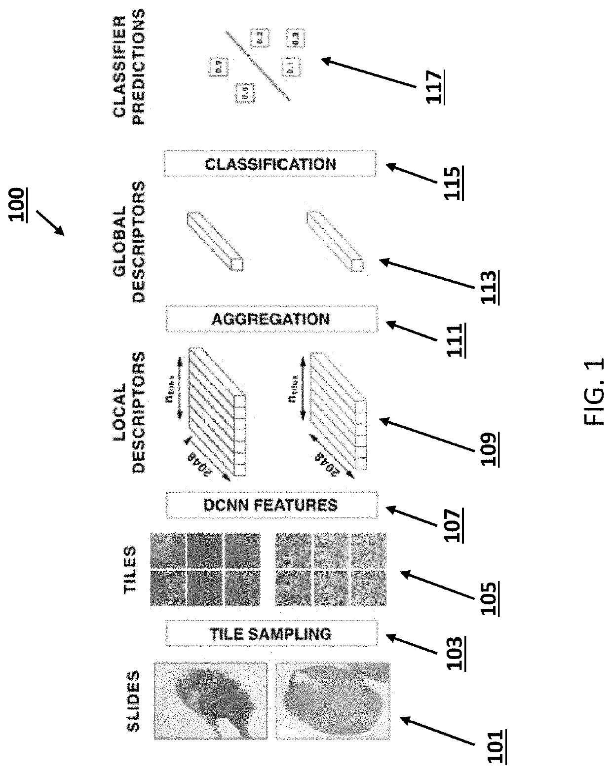
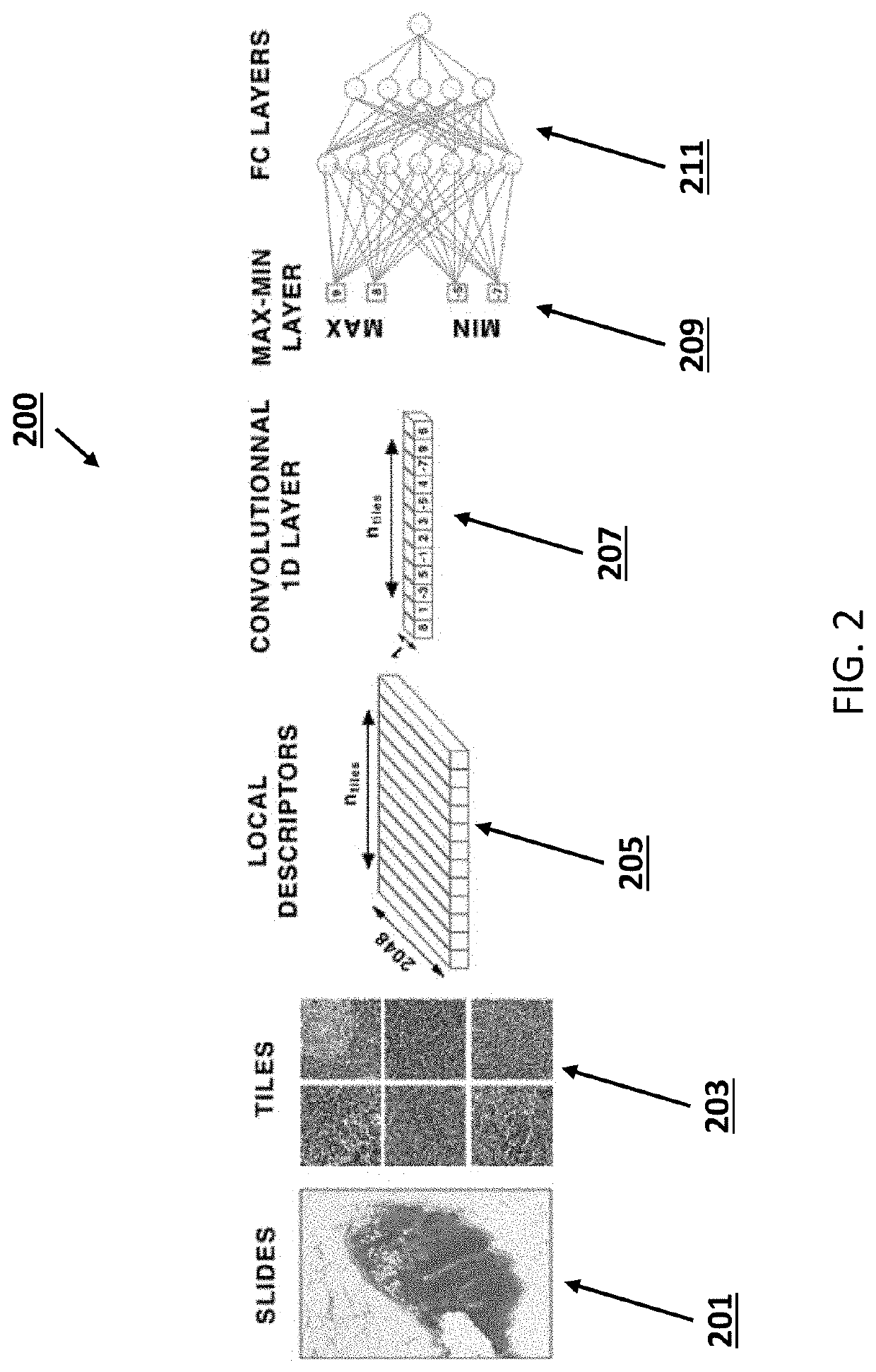
The patent describes a method and apparatus for classifying and predicting the global score of an image using a set of tiles. Each tile is extracted from the image and processed using a convolutional neural network to create a feature vector. These feature vectors are then used to calculate a score for each tile, which is used to classify the image. The method can be applied to histopathology slides and can predict the risk score or response to treatment. The technical effect of this invention is to provide a more accurate and efficient way to classify and predict the global score of images, which can be useful in various fields such as medical diagnosis.

Problems solved by technology

These localized annotations are even more costly to obtain than the original diagnosis, because pathologists must spend time to assemble pixel-by-pixel segmentation maps of diseased tissue at extreme resolution.
Thus, HIA datasets with annotations are very limited in size.
Additionally, such localized annotations may not be available when facing new problems in HIA, such as new disease subtype classification, prognosis estimation, or drug response prediction.
Indeed, while histopathology is the gold standard diagnostic in oncology and other disorders, it is extremely costly, requiring many hours of focus from pathologists to make a single diagnosis.
Additionally, as correct diagnosis for certain diseases requires pathologists to identify a few cells out of millions, these tasks are akin to “finding a needle in a haystack.” Hard numbers on diagnostic error rates in histopathology are difficult to obtain, being dependent upon the disease and tissue in question as well as self-reporting by pathologists of diagnostic errors.
However, false negatives in cancer diagnosis can lead not only to catastrophic consequences for the patient, but also to incredible financial risk to the pathologist.
Medical researchers have looked to computer-aided diagnosis for decades, but the lack of computational resources and data have prevented widespread implementation and usage of such tools.

Method used


Image

  • Systems and methods for image classification
  • Systems and methods for image classification
  • Systems and methods for image classification

Examples


examples

[0053]In one embodiment, for pre-processing, a single tile scale was fixed for all methods and datasets. A fixed zoom level of 0.5 μm / pixel was chosen, which corresponds to l=0 for slides scanned at 20× magnification, or l=1 slides scanned at 40× magnification. Next, since WSI datasets often contain a few hundred images, far from the millions images of ImageNet dataset, strong regularization was required to prevent over-fitting. A l2-regularization of 0.5 was applied on the convolutional feature embedding layer, and dropout on the MLP with a rate of 0.5. However, these values may not be the global optimal, as no hyper-parameter optimization was applied to tune these values. In some embodiments, the model parameters may be optimized to minimize the binary cross-entropy loss over 30 epochs with a mini-batch size of 10 and with learning rate of 0.001.

[0054]To reduce variance and prevent over-fitting, an ensemble of E CHOWDER networks was trained which differ by their initial weights. T...


PUM


Abstract

A method and apparatus of a device that classifies an image is described. In an exemplary embodiment, the method includes tiling at least one region of interest of the input image into a set of tiles. For each tile, the method includes extracting a feature vector of the tile by applying a convolutional neural network, wherein a feature is a local descriptor of the tile; and computing a score of the tile from the extracted feature vector, said tile score being representative of a contribution of the tile into a classification of the input image. The method also includes sorting a set of the tile scores and selecting a subset of the tile scores based on their value and / or their rank in the sorted set. The method also includes applying a classifier to the selected tile scores in order to classify the input image

Description

RELATED APPLICATION[0001]This Application claims priority and the benefit of U.S. Provisional Patent Application 62 / 799936 titled “CLASSIFICATION AND DISEASE LOCALIZATION IN HISTOPATHOLOGY USING ONLY GLOBAL LABELS: A WEAKLY-SUPERVISED APPROACH” filed on Feb. 1, 2019, the entire contents of which are incorporated herein by reference.FIELD OF INVENTION[0002]This invention relates generally to machine learning and computer vision and more particularly to image processing and classification.BACKGROUND OF THE INVENTION[0003]Histopathological image analysis (HIA) is a critical element of diagnosis in many areas of medicine, and especially in oncology, where this type of image analysis currently defines a gold standard metric. Recent works have sought to leverage modern developments in machine learning (ML) to aid pathologists in disease detection tasks, but the majority of these techniques require localized annotation masks as training data. These localized annotations are even more costl...

Claims


Application Information

Patent Timeline
Patent Type & Authority Applications(United States)
IPC IPC(8): G06K9/00G06K9/62G06K9/46G06T7/00G06V10/764
CPCG06K9/6261G06K9/00147G06T2207/30024G06T7/0012G06K9/4642G06T2207/20084G06K9/0014G06T2207/10024G06T2207/20021G06T2207/10056G06T2207/20081G06V2201/03G06V10/82G06V10/764G06V20/698G06V20/695G06F18/2163
Inventor COURTIOL, PIERRETRAMEL, ERIC W.SANSELME, MARCWAINRIB, GILLES
Owner OWKIN INC
Learn More