Methods and Apparatuses for Assessment and Management of Hemodynamic Status

a technology of hemodynamic status and hemodynamic status, applied in the field of hemodynamic status assessment and management, can solve the problems of non-ventilation patient history failure, non-ventilation patient current system cannot be used, so as to reduce arterial pressure, reduce the effect of arterial pressure, and convenient and cost-effectiveness

Active Publication Date: 2020-09-17
MEDICI TECH LLC
View PDF0 Cites 8 Cited by
  • Summary
  • Abstract
  • Description
  • Claims
  • Application Information

AI Technical Summary

Benefits of technology

[0017]The present invention is related to U.S. provisional applications 62 / 263,839, 62 / 375,431, and 62 / 423,701, each of which is incorporated herein by reference. Embodiments of the present invention provide reliable, convenient, and cost-effective methods and apparatuses to determine the hemodynamic status of the patent. The methods and apparatuses provide for the noninvasive determine of hemodynamic status by using systematic perturbations of venous return or trend observation over time. Embodiments do not require invasive pressure monitoring or the use of ventilator but instead can be an entirely noninvasive system. The present invention represents a departure from prior approaches by using mini-Mueller and mini-Valsalva controlled breathing activities or patient-initiated changes in body position to create systematic changes in venous return. These changes in cardiac function are then evaluated on a beat-to-beat basis with awareness of the phase of the breathing (inhale or exhale), the position of the patient, or both. The system provides important information on fluid responsiveness including dynamic and static parameters for hemodynamic assessment for treatment of patients in the emergency department and urgent care, the dialysis clinic, as well as the home of the patient. Embodiments of the invention allow the care provider to determine if blood flow to the body will increase (or decrease) if the patient's intravascular volume is increased (or decreased), and whether any decrease in arterial pressure is due to loss of vascular tone or due to inadequate blood flow. Additionally, the system is able to determine the onset of hemodynamic congestion prior to the development of physical symptoms and to characterize the degree of heart failure through analysis of the subject's response to a systematic perturbation in venous return.

Problems solved by technology

Although there are a few publications showing positive results the overwhelming consensus is that spontaneous breathing does not work with current processing methods.
No existing system can be used on non-ventilated patients.
Pinsky explains the problems well by stating that historical efforts have failed in non-ventilated patients due to a variety of factors that explain the lack of performance.
Spontaneous inspiratory efforts can increase intra-abdominal pressure because of active compression of abdominal muscles, exaggerating the preload response.
Pinsky also discussed limitation of the standard Valsalva maneuver because the Valsalva maneuver can also affect right and left ventricular afterload, which can contribute to respiratory variations in stroke volume.
Pinsky concludes his review of issues by stating “regrettably, ΔPP and other derived indices cannot be used in spontaneously breathing patients, as slight and sometimes undetected changes in breathing pattern may affect these variables”.
Muller at al. states that dynamic indices such as arterial pulse pressure or aortic velocities recorded by esophageal Doppler or echocardiography are not valid in spontaneously breathing patients.
However, the authors recommend caution as low variations (<40%) of IVC diameter cannot rule out a need for fluid therapy in spontaneously breathing patients with acute circulatory failure.
However, the performance during this maneuver was significantly lower than during quiet respiration.
The publication concludes that their findings confirm the poor value of clinical signs and / or standard hemodynamic parameters to predict the effects of fluid expansion in spontaneous breathing patients.
The results suggest that pulse pressure variation and systolic pressure variation are less effective in predicting fluid responsiveness during spontaneous breathing than in mechanical ventilation.
Although Soubrier at al. demonstrated decreased performance with forced exhale, other authors have shown improved results in limited testing.
The publication has similar limitations to Bronzwaer but the influence of anesthesia and the use of pigs makes the study hard to translate to ambulatory, awake humans.
The protocol and system used do not address the issues defined by Pinsky in terms of a creating a repeatable test as the degree of inspiration is not controlled.
Additionally, the length of the Valsalva at 10 second will be difficult for many patients to perform.
In these cases, static markers of cardiac preload are generally in the normal range and are rarely helpful for determination of volume responsiveness.
Because of the presence of spontaneous breathing, the indices of volume responsiveness that use heart-lung interactions, such as respiratory variation in arterial pressure and in stroke volume are no longer reliable.”
In the discussion, the authors site possible issues with normalization of heart rate, possible problems with the breathing protocol, and the degree of change in the Frank-starling curve.
Specifically, the authors state that “a main limitation of using LVETp and PTT as indicators of blood loss is their relatively large inter-subject variations in comparison with the changes induced by blood donation, which means that it is their intra-subject changes rather than their absolute values that are clinically useful, as with most other hemodynamic measurements.
In addition, significant increases in AMBR were found due to blood loss in ear and finger PPG signals.
Testing was limited to healthy individuals with a narrow age distribution.
A limitation of prior work has been the inability to determine the position of the finger- or head-based PPG sensors, specifically the sensor's vertical position relative to the heart.
Historically, the processes used electrodes placed on the skin, but newer devices no longer use electrodes.
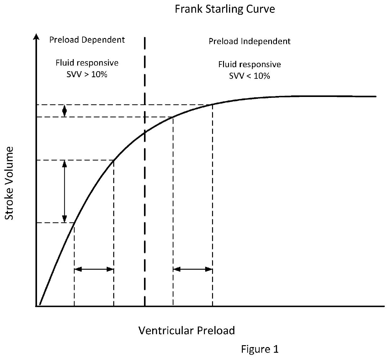
The resulting cardiac operating points cause in a large change in the cardiac output or stroke volume.
The ability to simulate this define movement without a mechanical bed is almost impossible.
Examination of FIG. 9 illustrates, that inspiration due to decreased lower intrathoracic pressure can result in venous collapse especially under hypovolemic conditions.
The result is a damped response to right heart changes.
These changes will have a longer time constant and this difference in timing can be significant when processing the measured signals.
A single Valsalva maneuver is a single measurement and is difficult for the subject to complete due to the duration of the maneuver and the moderately high intrathoracic pressures.
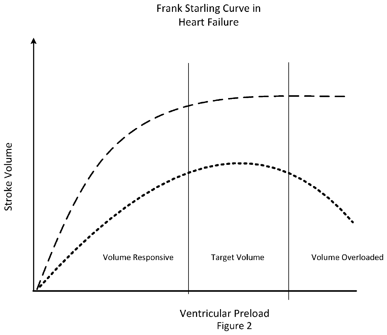
This phenomenon becomes the predominant component of stroke volume fluctuations during hypervolemia and / or congestive heart failure and is not predictive of fluid responsiveness.
The fact that conventional metrics of systolic pressure variance and stroke volume variance are based on the difference between the maximal and minimal values of systolic arterial pressure during the mechanical breath might reduce their accuracy in the prediction of volume responsiveness, especially in the presence of impaired left ventricular function.
In stark contrast, decreased intrathoracic pressure can result in venous collapse and a plateau in terms of maximal venous return.
First, as noted above, the response time associated with ventricular interdependence effects is rapid due to the lack of any air damping.
However, a sizable change in the volume of the right ventricle leads to a decrease in the volume of the left ventricle because of a leftward shift of the interventricular septum.
In fact, systolic overload of the right ventricle results in the most severe geometric configurational changes of the left ventricle.

Method used


Image

  • Methods and Apparatuses for Assessment and Management of Hemodynamic Status
  • Methods and Apparatuses for Assessment and Management of Hemodynamic Status
  • Methods and Apparatuses for Assessment and Management of Hemodynamic Status

Examples


example embodiments

[0240]First Example Embodiment and Operation. The example method and apparatus creates a simple test that enables determination of hemodynamic status and subsequent determination of appropriate treatments for subjects that might be hemodynamically compromised. The device shown in

[0241]FIG. 41 includes the following measurement systems:

A ECG measurement system, for example from the right thumb

A PPG measurement system, for example on the index finger of the patient,

A PPG measurement system on the bottom of the device for chest PPG measurements; and

A PCG measurement system located on the bottom of the device

[0242]The device as pictured can resemble the typical computer mouse in terms of size and ergonomics. The patient can place the device over the heart as shown in FIG. 43. The system can ensure that a quality phonocardiogram signal is obtained and provide feedback to the user regarding correct placement of the device. The system can also assess the quality of both the ECG and PPG sig...


PUM


Abstract

Embodiments of the present invention provide reliable, convenient, and cost-effective methods and apparatuses to determine the hemodynamic status of the patent. The methods and apparatuses provide for the noninvasive determine of hemodynamic status by using systematic perturbations of venous return or trend observation over time. Embodiments do not require invasive pressure monitoring or the use of ventilator but instead can be an entirely noninvasive system.

Description

BACKGROUND OF THE INVENTION[0001]The determination of hemodynamic status and subsequent treatment of hemodynamically compromised patients is challenging. Treatment decisions are often based upon the performance of two critical physiological processes: cardiac function and venous return function, which is determined by the return of blood to the heart. The management of cardiac output is dependent upon changes to one or both of these two functions. Pinsky has simplified the complex therapeutic decision process to two functional performance-based questions: (1) Will blood flow to the body increase (or decrease) if the patient's intravascular volume is increased (or decreased), and if so, by how much, and (2) Is any decrease in arterial pressure due to loss of vascular tone or merely due to inadequate blood flow? These two questions can be summarized as the determination of fluid responsiveness and the determination of vascular tone, respectively.[0002]Various publications are referenc...

Claims


Application Information

Patent Timeline
Patent Type & Authority Applications(United States)
IPC IPC(8): A61B5/0205A61B5/024A61B5/02A61B5/029A61B5/091A61B5/00
CPCA61B5/0024A61B5/0205A61B5/02028A61B5/029A61B5/02405A61B5/091A61B5/02125A61B5/02416A61B5/1116A61B5/113A61B5/4561A61B7/00A61B5/4812A61B5/1118A61B5/6826A61B5/349A61B5/02427A61B5/1126
Inventor ROBINSON, MARK RIESALLEN, ELENA A
Owner MEDICI TECH LLC
Why Eureka
Try Eureka