Image contrast enhancement of an x-ray image

a technology of contrast enhancement and x-ray image, which is applied in image enhancement, angiography, instruments, etc., can solve the problems of insufficient contrast enhancement and achieve the effect of improving and stabilizing contrast enhancemen

Active Publication Date: 2021-05-20
KONINKLJIJKE PHILIPS NV
View PDF0 Cites 2 Cited by
  • Summary
  • Abstract
  • Description
  • Claims
  • Application Information

AI Technical Summary

Benefits of technology

[0024]As a result, improved and stable contrast enhancement applied to X-ray images can be achieved, e.g. optimized opacification and delineation in the anatomy, including lesions, can be achieved.
[0035]According to an aspect, it is provided to enhance the image quality of an acquired X-ray image by adapting contrast boosting or contrast enhancement depending on the circumstances of the acquired image. As a result, the contrast enhancement is done dynamically. For example, when an image is captured for a thicker patient, the adapted contrast enhancement parameter is adapted to a higher level than for a thinner patient.

Problems solved by technology

However, it has been shown that contrast enhancement is still not always leading to sufficient results.

Method used


Image

  • Image contrast enhancement of an x-ray image
  • Image contrast enhancement of an x-ray image

Examples


Embodiment Construction

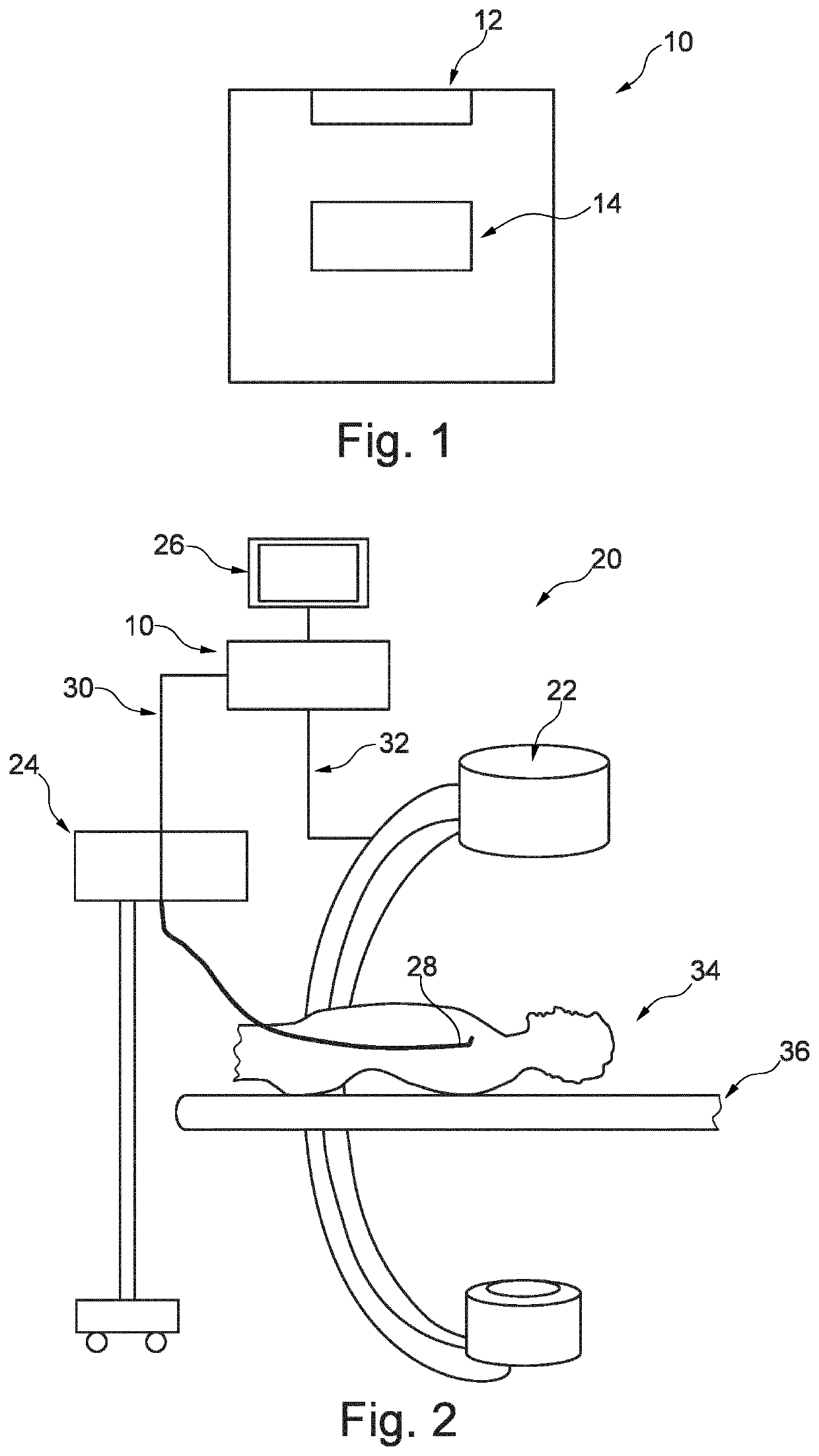
[0042]FIG. 1 shows a device 10 for image contrast enhancement of an X-ray image of a vascular structure. The device 10 comprises an input unit 12 and a processing unit 14. The input unit 12 is configured to provide an acquired X-ray image of a vascular structure with a contrast injection (as shown in FIG. 2). The contrast injection is performed with a current contrast injection setting having at least one contrast injection parameter. The input unit 12 is configured to provide a generic vascular structure. The input unit 12 is also configured to provide the current contrast injection setting. The processing unit 14 is configured to determine an assessed contrast parameter for the generic vascular structure based on the current contrast injection setting. The processing unit 14 is also configured to determine an adapted image contrast enhancement for the generic vascular structure based on the assessed contrast parameter. The processing unit 14 is further configured to apply the adap...


PUM


Abstract

The present invention relates to image contrast enhancement. In order to provide improved and stable contrast enhancement for each acquired image, a device (10) for image contrast enhancement of an X-ray image of a vascular structure is provided. The device (10) comprises an input unit (12) and a processing unit (14). The input unit is configured to provide an acquired X-ray image of a vascular structure with a contrast injection. The contrast injection is performed with a current contrast injection setting having at least one contrast injection parameter. The input unit is further configured to provide a generic vascular structure. The input unit is also configured to provide the current contrast injection setting. The processing unit is configured to determine an assessed contrast parameter for the generic vascular structure based on the current contrast injection setting. The processing unit is further configured to determine an adapted image contrast enhancement for the generic vascular structure based on the assessed contrast parameter. The processing unit is also configured to apply the adapted image contrast enhancement to the acquired X-ray image in order to generate a contrast-enhanced X-ray image.

Description

FIELD OF THE INVENTION[0001]The present invention relates to a device for image contrast enhancement of an X-ray image of a vascular structure, to a system with a such device, to a method for image contrast enhancement of an X-ray image of a vascular structure, to a computer program element for controlling such a device and to a computer readable medium having stored the program element.BACKGROUND OF THE INVENTION[0002]In X-ray images, contrast enhancement (CE) is provided to improve image quality for the viewer. Contrast enhancement may be realized in the framework of frequency decomposition. For example, the image is decomposed into a certain number of frequency sub-bands, for instance, with a so-called Laplacian Pyramid. The anatomical or interventional details one is willing to enhance are spread across several of those frequency sub-bands. Gains, which may depend on the local intensity are then applied to the content of those sub-bands, before the total spectrum is re-composed,...

Claims


Application Information

Patent Timeline
Patent Type & Authority Applications(United States)
IPC IPC(8): A61B6/00G06T5/00G16H30/40
CPCA61B6/5205G06T5/007G06T2207/30101G06T2207/10116G16H30/40A61B6/481A61B6/504A61B6/5211A61B6/5258A61B6/5294G16H50/20
Inventor HOORNAERT, BART PIERRE ANTOINE JOZEFFLORENT, RAOULLEVRIER, CLAIREGEVERS, BERNARDUS MARIUS HUBERTUS
Owner KONINKLJIJKE PHILIPS NV
Why Eureka
Try Eureka