Reading codes via a surgical microscope camera

a technology of barcode scanner and code reader, which is applied in the field of surgical microscope operation, can solve the problems of virtually impossible for technicians or service staff to know these numbers, time-consuming and easy errors, and inability to read codes via user interface, and achieve the effect of facilitating the use of codes and ensuring reliability

Pending Publication Date: 2021-09-30
CARL ZEISS MEDITEC AG
View PDF4 Cites 0 Cited by
  • Summary
  • Abstract
  • Description
  • Claims
  • Application Information

AI Technical Summary

Benefits of technology

[0008]It is an object of the invention to provide an advantageous method for operating a surgical microscope, as a result of which, in particular, the use of codes is facilitated in simpler and, at the same time, more reliable fashion.

Problems solved by technology

As a rule, the manual input of codes, passwords, and serial numbers via a user interface is time-consuming and susceptible to errors.
It is virtually impossible for the technician or the member of servicing staff to know these numbers off by heart since each device is provided with individual codes and passwords.
As a result, scanning static codes using conventional barcode scanners is no longer a suitable option.
It is usually not possible to automatically log the performance.
This system is complicated and expensive since individual hardware, specifically the RFID reader, must be installed in this case.
This significantly increases the production costs of the device.
The RFID target on the drape also leads to higher production costs, albeit only to a relatively small increase.
Moreover, an RFID reader also means significantly increased complexity within the scope of the approval of the device since the reading method uses radio waves which require a radio license and possibly an EMC test.

Method used


Image

  • Reading codes via a surgical microscope camera

Examples


Embodiment Construction

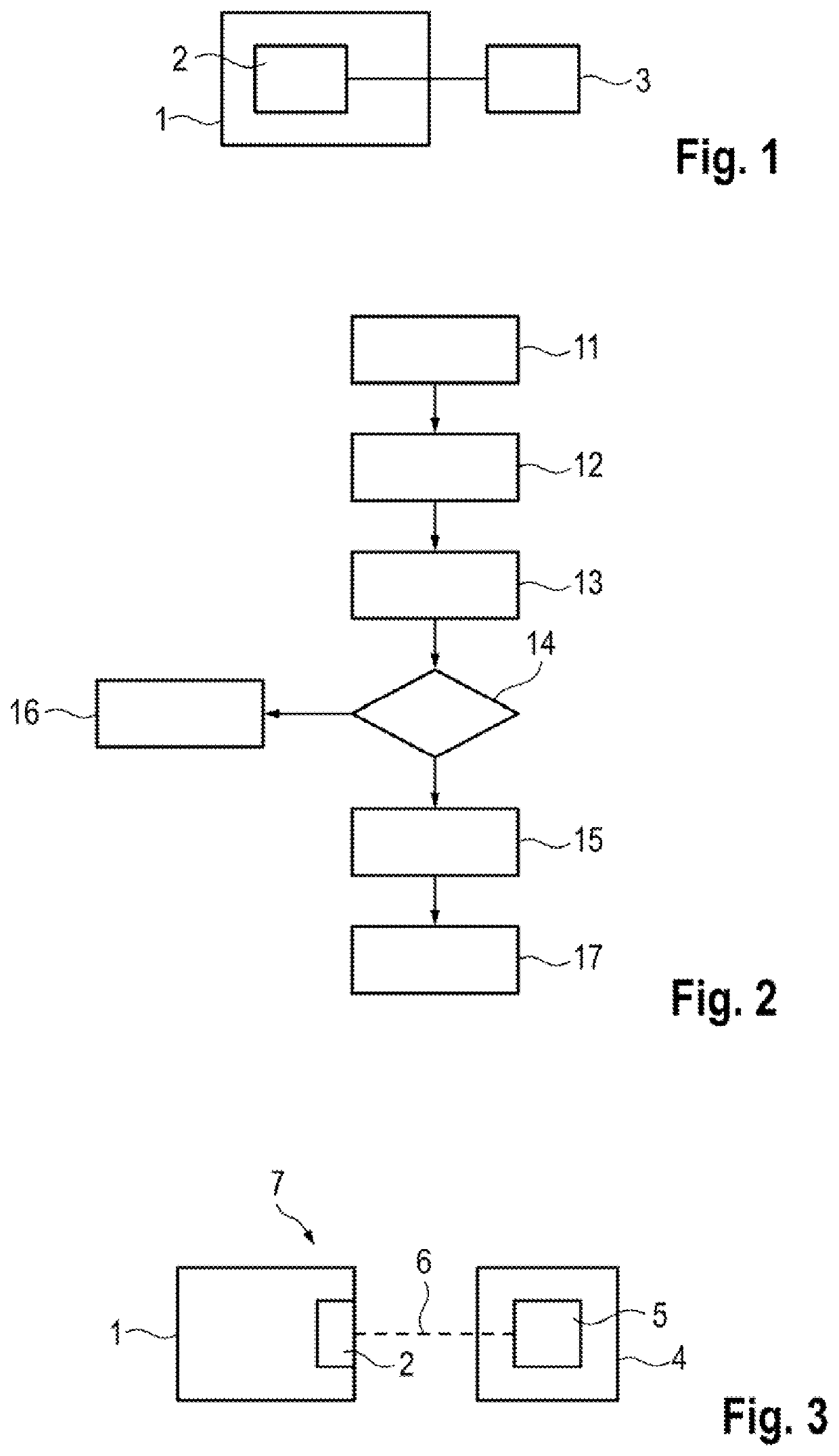
[0034]FIG. 1 schematically shows a surgical microscope 1. The surgical microscope 1 includes a camera 2. The surgical microscope 1 is configured to be operated in accordance with an embodiment described in exemplary fashion below on the basis of FIG. 2. To this end, the camera 2 is connected to an evaluation device 3 for data transmission purposes. The evaluation device 3 can be a constituent part of the surgical microscope or can be configured as a separate apparatus, for example, as a computer connected to the surgical microscope.

[0035]FIG. 2 schematically shows an example for a method according to the disclosure in the form of a flowchart. In a first step 11, a code that is able to be captured in a set frequency range, for example a visually perceivable or imaged code, is introduced into the field of view of the camera 2. To this end, the code can be guided into the field of view of the camera and / or the field of view of the camera can be directed at the code. By way of example, ...


PUM


Abstract

A method for operating a surgical microscope having a camera is described. The method includes the following steps: a code that is able to be captured in a set frequency range is introduced into the field of view of the camera, at least one image which includes the code is captured via the camera, an evaluation device is used to identify the code and compare the latter to set features of a code required to activate at least one function, and if the identified code has the set features, the activation of the function is authorized.

Description

CROSS REFERENCE TO RELATED APPLICATION[0001]This application claims priority of German patent application no. 10 2020 108 345.9, filed Mar. 26, 2020, the entire content of which is incorporated herein by reference.FIELD OF THE INVENTION[0002]The disclosure relates to a method for operating a surgical microscope including a camera, in particular having a camera integrated into the surgical microscope. Moreover, the disclosure relates to a surgical microscope and an arrangement for performing a surgery.BACKGROUND OF THE INVENTION[0003]The amount of electronics and software used in modern surgical microscopes is steadily increasing. Prior to surgery, the users must frequently test various functions in order to check the function of the device and ensure greater patient safety. To this end, it is necessary to activate various menus throughout the test. In this respect, it is necessary to perform various entries on the user interface (GUI—graphical user interface). By way of example, it ...

Claims


Application Information

Patent Timeline
Patent Type & Authority Applications(United States)
IPC IPC(8): G02B21/00G02B21/36A61B90/00A61B90/96
CPCG02B21/0012A61B90/96A61B90/361G02B21/361A61B90/20A61B90/94
Inventor BART, FLORIANLORENZ, AXELKOENIG, FRANKKEIB, FRANK
Owner CARL ZEISS MEDITEC AG
Why Eureka
Try Eureka