Imaging method using pain sensor

a technology of pain sensor and imaging method, which is applied in the field of supporting imaging and imaging arrangement, can solve the problems of pain in the patient's fixation of mamma or other body parts, breast cancer remains a leading cause of death in women, and achieves the effects of improving contrast sensitivity and specificity, reducing pain and hence stress level, and improving image quality

Pending Publication Date: 2021-10-14
KONINKLJIJKE PHILIPS NV
View PDF0 Cites 0 Cited by
  • Summary
  • Abstract
  • Description
  • Claims
  • Application Information

AI Technical Summary

Benefits of technology

[0055]The fixation device allows exerting different levels of physical pressure, and thus compression, to improve contrast sensitivity and specificity.
[0056]The proposed system allows overcoming to red

Problems solved by technology

Breast cancer remains a leading cause of death in women.
Fixation of the mamma, or

Method used


Image

  • Imaging method using pain sensor
  • Imaging method using pain sensor
  • Imaging method using pain sensor

Examples


Embodiment Construction

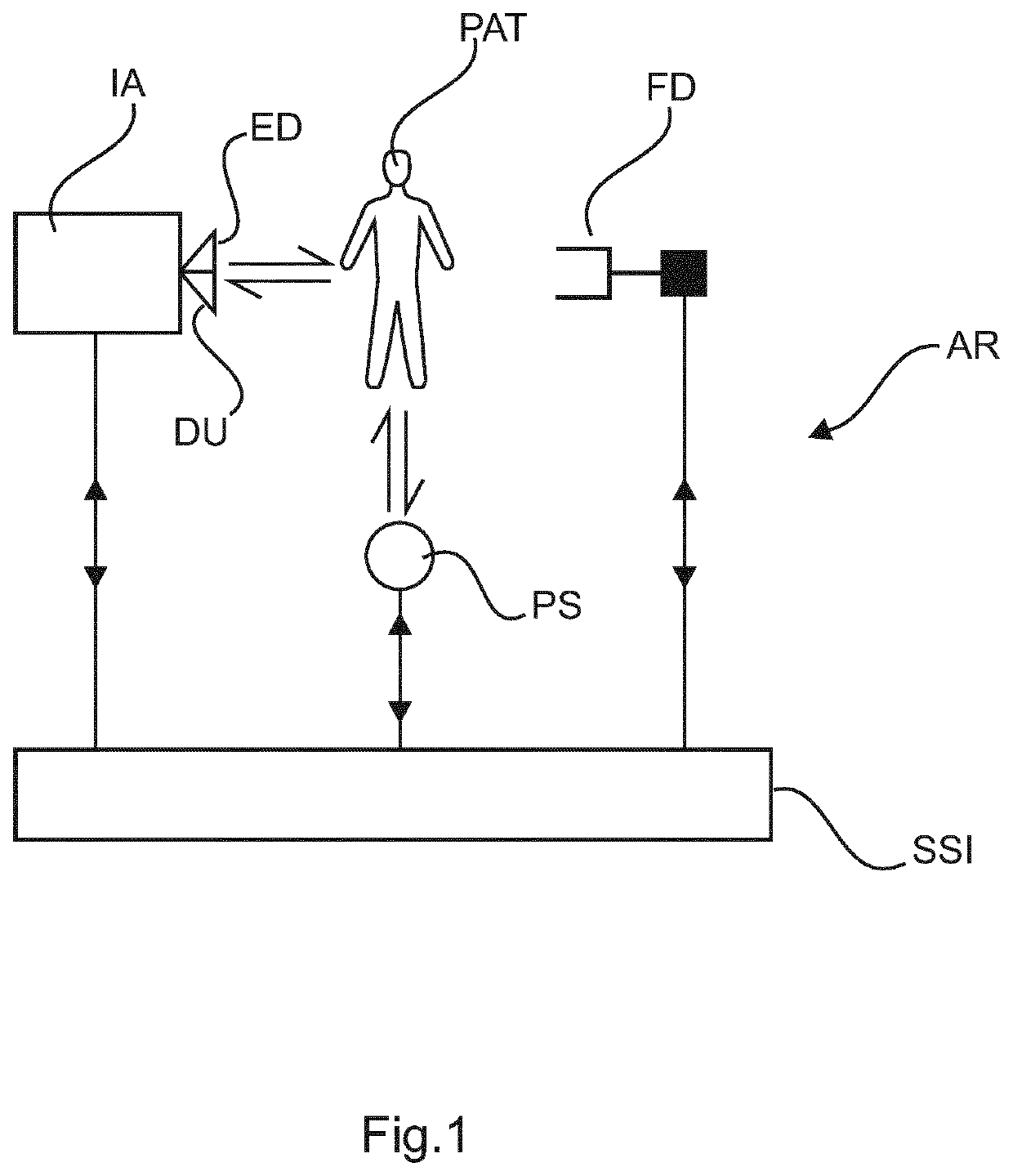
[0074]Referring first to FIG. 1, this shows a schematic block diagram of an imaging arrangement AR as envisaged herein in embodiments. The imaging arrangement AR includes an imaging apparatus IA configured to acquire medical images of a human or animal patient PAT. Different modalities are envisaged herein including, preferably MRI, but also x-ray, emission imaging such as PET or SPECT, or ultrasound imaging are all envisaged herein.

[0075]In operation, the imaging apparatus IA performs an imaging procedure. During the imaging procedure the patient PAT is exposed to an interrogating signal such as ionizing or non-ionizing radiation emitted from an optional emitter device ED. In emission imaging such as PET, there is no emitter device ED. In this case the interrogating signal is emitted by a radioactive tracer substance from within the patient, the substance having been previously administered to patient PAT.

[0076]Exposure to the interrogating signal may be restricted to a certain reg...


PUM


Abstract

A system for controlling operation of an imaging or therapy apparatus. The system comprises an interface (IN) for receiving a pain measurement signal as measured by one or more sensors (S) in relation to an anatomic part (BR) of the patient (PAT) i) being imaged in an imaging procedure by the imaging apparatus or ii) being under therapy in a therapy procedure delivered by the therapy device, whilst the part (BR) is held in an adjustable fixation device (FD). A control unit (CU) of the system is configured to process the pain measurement signal to compute at least one control signal. A control interface (CIF) of the system is configured to interact during the imaging or therapy procedure with the imaging apparatus (IA) and/or the fixation device (FD) based on the control signal to a) influence the imaging or therapy procedure and/or b) to adjust the fixation device so as to change the manner in which the part (BR) is being held.

Description

FIELD OF THE INVENTION[0001]The invention relates to a system of supporting imaging, an imaging arrangement, to a method of supporting imaging, to a method of training a machine learning component, to a computer program element, to a computer readable medium, and to a fixation device.BACKGROUND OF THE INVENTION[0002]Breast cancer remains a leading cause of death in women.[0003]Overall, mammography is considered the single most effective screening tool and has been credited with reducing breast cancer-related mortality.[0004]There is no doubt that mammography is a very cost-effective tool for breast cancer screening. Breast MRI is currently the most sensitive detection technique for breast cancer diagnosis. It is able to detect cancer not visible on conventional imaging (such as X-ray based), it can be used as a problem-solving instrument, and it can be applied to screen high-risk patients. Breast MRI is also better at monitoring the response to chemotherapy than other imaging modali...

Claims


Application Information

Patent Timeline
IPC IPC(8): A61B5/00
CPCA61B5/4827A61B5/7267A61B5/708G16H20/10G16H20/40G16H30/20G16H40/63
Inventor LEUSSLER, CHRISTOPHHELLE, MICHAEL GÜNTERWIRTZ, DANIELVOGTMEIER, GEREONJOHNSON, MARK THOMAS
Owner KONINKLJIJKE PHILIPS NV
Why Eureka
Try Eureka