Methods for Automated Lesion Analysis in Longitudinal Volumetric Medical Image Studies

a volumetric medical image and automated technology, applied in the field of image analysis, can solve the problems of significant intra- and inter-observer variability, and the delineation of manual lesions for the purpose of volumetric measurement is rarely performed in the clinic today

Inactive Publication Date: 2021-10-21
YISSUM RES DEV CO OF THE HEBREWUNIVERSITY OF JERUSALEM LTD +1
View PDF2 Cites 3 Cited by
  • Summary
  • Abstract
  • Description
  • Claims
  • Application Information

AI Technical Summary

Benefits of technology

[0028]2) simultaneous, coupled detection in two or more images.
[0029]For purposes of this specification and the accompanying claims, the term “registration” indicates alignment of two of more images so that they have the same coordinate system using 3D rigid and deformable registration (Group-wise image registration based on a total correlation dissimilarity measure for quantitative MRI and dynamic imaging data. Guyader et al, Nature Scientific Reports, 2018.)

Problems solved by technology

However, volumetric measurements require delineating the lesion contours in the scan slices, which is a tedious, time-consuming, and error prone task that requires medical expertise and that may yield significant intra- and inter-observer variability.
Consequently, manual lesions delineation for the purpose of obtaining volumetric measurements is seldom performed in the clinic today.

Method used


Image

  • Methods for Automated Lesion Analysis in Longitudinal Volumetric Medical Image Studies
  • Methods for Automated Lesion Analysis in Longitudinal Volumetric Medical Image Studies
  • Methods for Automated Lesion Analysis in Longitudinal Volumetric Medical Image Studies

Examples


Embodiment Construction

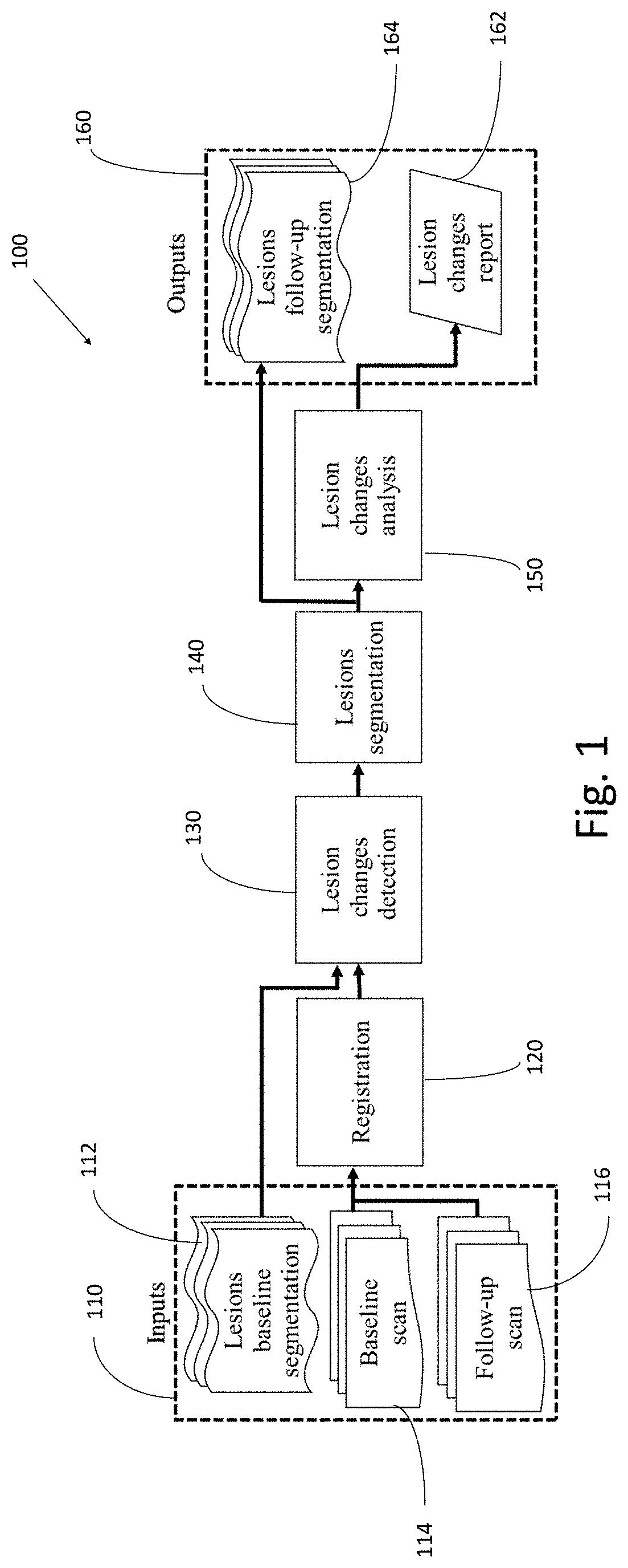
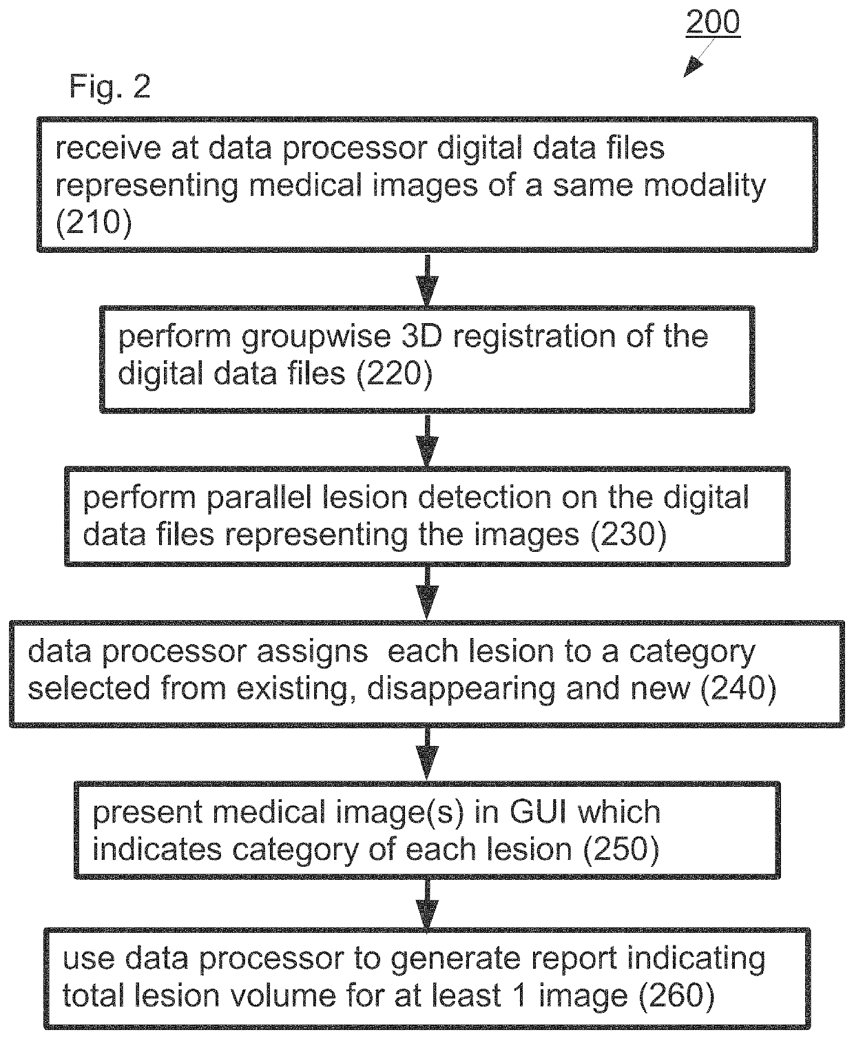
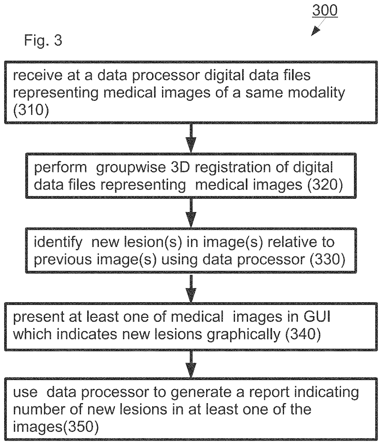
[0042]Embodiments of the invention relate to methods for automation of radiological follow up as well as user interfaces that present findings graphically.

[0043]Specifically, some embodiments of the invention can be used to determine for each tumor in a patient whether it is new, previously existing or has disappeared. In some embodiments, change in characteristics of previously existing tumors is automatically determined (i.e. amount of growth / shrinkage). Alternatively or additionally, in some embodiments a quantitative lesion and / or lesion changes report is generated automatically.

[0044]The principles and operation of a methods and / or graphical user interfaces (GUIs) according to exemplary embodiments of the invention may be better understood with reference to the drawings and accompanying descriptions.

[0045]Before explaining at least one embodiment of the invention in detail, it is to be understood that the invention is not limited in its application to the details set forth in t...


PUM


Abstract

Described herein is a computer implemented method that includes receiving at a data processor two or more digital data files representing medical images of a same modality; performing group-wise 3D registration of the digital data files representing medical images of a same modality; and parallel lesion detection and analysis on the digital data files representing the medical images.

Description

CROSS-REFERENCE TO RELATED APPLICATIONS[0001]Benefit is claimed to Israel Patent Application No. 274016, filed Apr. 18, 2020, the contents of which are incorporated by reference herein in their entirety.FIELD OF THE INVENTION[0002]The invention is in the field of image analysis.BACKGROUND OF THE INVENTION[0003]Radiology relies on medical images to diagnose and treat disease and is one of the cornerstones of modern healthcare. More than 1 billion volumetric scans (750 million CT and 200 million MRI) are performed annually in Europe, Asia, and the US. Currently each scan requires a radiologist to view it, analyze it, and to write a radiology report. Radiological follow-up is the interpretation of two or more radiological images acquired over the course of time to evaluate disease progression and to determine treatment efficacy in support of clinical decision-making Radiological follow-up is a key clinical task which is currently performed manually. Approximately 40% of expert radiolog...

Claims


Application Information

Patent Timeline
Patent Type & Authority Applications(United States)
IPC IPC(8): G06T7/00
CPCG06T7/0016G06T2200/24G06T2207/20081G06T2207/20084G06T2207/30096G06T7/0012G06T7/30G06T7/11
Inventor JOSKOWICZ, LEOSOSNA, JACOB
Owner YISSUM RES DEV CO OF THE HEBREWUNIVERSITY OF JERUSALEM LTD
Why Eureka
Try Eureka