A method for determining the likelihood of a patient being responsive to cancer immunotherapy
a cancer immunotherapy and patient technology, applied in the field of determining the likelihood of a patient being responsive to cancer immunotherapy, can solve the problems of cancer death among women worldwide and lack of understanding of the breast cancer ecosystem, and achieve the effect of high likelihood of being
- Summary
- Abstract
- Description
- Claims
- Application Information
AI Technical Summary
Benefits of technology
Problems solved by technology
Method used
Image

Examples
example 1
Cell Proteomic Atlas of Breast Cancer Ecosystems
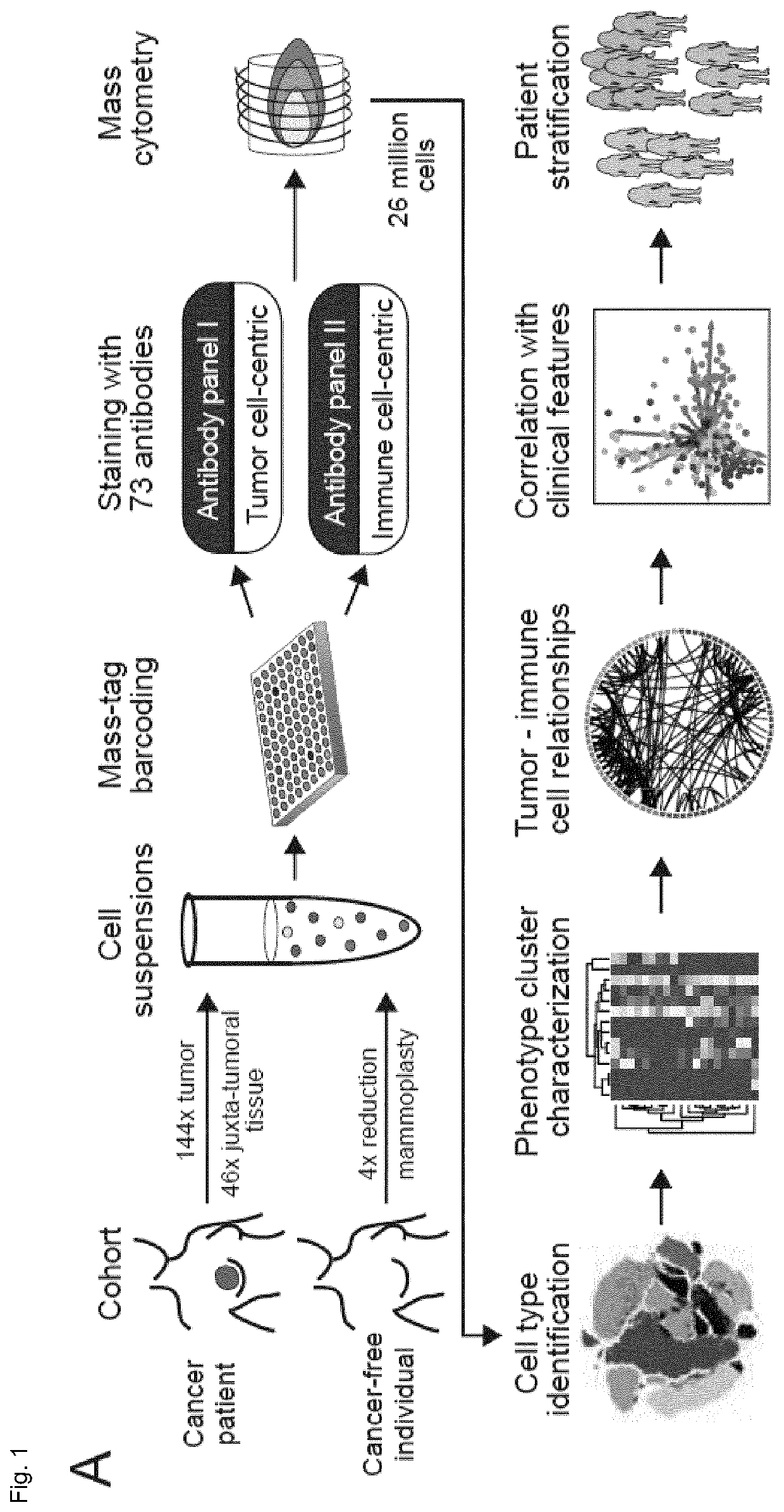
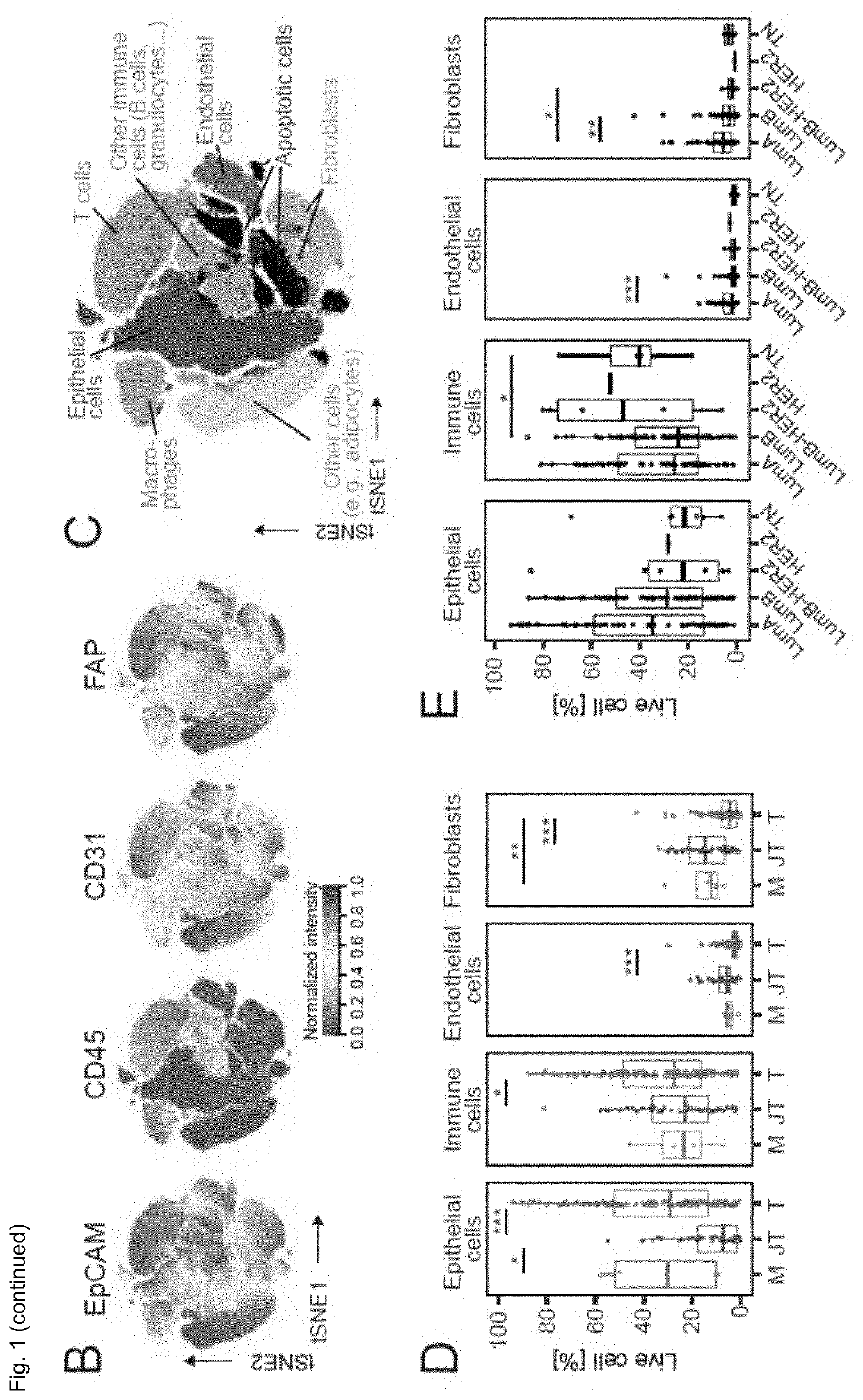
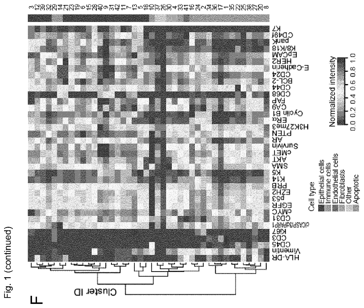
[0122]The inventors performed large-scale mass cytometry profiling of 144 prospectively collected tumors, including 56 Luminal A, 72 Luminal B, six Luminal B-HER2+, one HER2+ and six TN tumors (Table 1) (Coates et al., 2015, Ann. Oncol. 26, 1533-1546). Histopathology divided the samples into 106 invasive ductal, 16 invasive lobular, and 22 mixed / other tumors. An automated system was used to generate single-cell suspensions from all tissue samples (STAR Methods). These samples and seven breast cancer cell lines were mass-tag barcoded (Zunder et al., 2015, Nat. Protoc. 10, 316-333), pooled for antibody staining with 73 antibodies, and simultaneously analyzed by mass cytometry (FIG. 1A). One antibody panel was tumor-cell centric to quantify markers that identify mammary cell types, signaling, proliferation, and survival. The other panel was focused on immune phenotyping and based on the inventors' recent immune cell atlas of clear cell re...
example 2
e Landscape of Breast Cancer
[0125]T cells and myeloid cells were the most abundant immune cell types in the inventors' study; fewer natural killer (NK) cells, B cells, granulocytes, plasma cells, and plasmacytoid dendritic cells were detected (FIGS. 2A, S2A-D). Breast tumors were enriched for T cells and B cells and contained a lower frequency of NK cells and granulocytes than juxta-tumoral tissue (FIG. 2A). There was considerable inter-patient variation in tumor-associated immune cell frequencies (FIG. 2A) as previously described.
[0126]T cells and macrophages can exert pro-tumor or anti-tumor activities. In-depth analyses of T cells by t-SNE and PhenoGraph identified ten CD4+ and ten CD8+ T cell clusters (T01-T20; FIGS. 2B-D). Most T cell clusters had an effector memory phenotype (CD197+, CCR7low, CD45RAlow), and tumor-associated T cells existed as a phenotypic continuum across the CD4+ and CD8+ lineages (FIG. 2D). Various levels of PD-1 and heterogeneous co-expression of co-inhibi...
example 3
mors are Enriched for Immunosuppressive Macrophage Phenotypes
[0130]To characterize TAM populations, t-SNE and PhenoGraph were applied to all myeloid cells (FIG. 2J), resulting in 19 myeloid clusters (M01-M19) of five categories: i) CD14-expressing classic (M06, CD14+CD16−) and inflammatory monocytes (M15, CD14intCD16+), ii) early immigrant macrophages (M03, M11, M13, HLA-DRintCD192+), iii) tissue-resident macrophages (M08, M09, M16, CD206+HLA-DRint), iv) TAMs (M01, M02, M04, M14, M17, CD64highHLA-DRhigh), and v) myeloid-derived suppressor cells (MDSCs; M07, M10, M12, HLA-DR-CD38+) (FIGS. 2K-L). Consistent with previous reports, the myeloid phenotypic space differed between tumor and juxta-tumoral regions (FIG. 2M). In 80% of tumors at least 10% of myeloid cells were PD-L1+. The PD-L1+ TAMs were phenotypically heterogeneous: TAMs in cluster M01 expressed pro-tumor markers CD204, CD206, CD163, and CD38 and anti-tumor marker CD169, whereas TAMs in M02 expressed CD204, CD169, and interm...
PUM
 Abstract
Description
Claims
Application Information
 - R&D
- Intellectual Property
- Life Sciences
- Materials
- Tech Scout
- Unparalleled Data Quality
- Higher Quality Content
- 60% Fewer Hallucinations
Browse by: Latest US Patents, China's latest patents, Technical Efficacy Thesaurus, Application Domain, Technology Topic, Popular Technical Reports.
© 2025 PatSnap. All rights reserved.Legal|Privacy policy|Modern Slavery Act Transparency Statement|Sitemap|About US| Contact US: help@patsnap.com