Unlock instant, AI-driven research and patent intelligence for your innovation.

Methods for Sacroiliac Joint Stabilization

a sacroiliac joint and joint technology, applied in the field of sacroiliac joint stabilization, can solve the problems of sacroiliac joint dysfunction, lower back pain associated with si joint dysfunction is suspected to be far more common, and surgeons are required to make a substantial incision in and through the skin

Pending Publication Date: 2022-09-22
TENON MEDICAL INC
View PDF0 Cites 3 Cited by
  • Summary
  • Abstract
  • Description
  • Claims
  • Application Information

AI Technical Summary

Benefits of technology

The present invention provides minimally-invasive methods for stabilizing dysfunctional joints in the spine, particularly the sacro-iliac joint. The methods involve using a tool assembly to access the joint and create a pilot opening for a prosthesis. A drill guide is then used to dislodge bone fragments and create a larger opening for the prosthesis. A prosthesis deployment assembly is then connected to the prosthesis and the assembly is guided into the joint. The invention offers a safer and less invasive method for stabilizing dysfunctional joints in the spine.

Problems solved by technology

As is also well established, the noted loads and, hence, forces exerted on the SI joint can adversely affect the biomechanical functions of the SI joint, which can, and often will, result in SI joint dysfunction—an often-overlooked musculoskeletal pathology associated with lower back pain.
However, lower back pain associated with SI joint dysfunction is suspected to be far more common than most healthcare providers realize, since such pain is often associated with other skeletal and musculoskeletal dysfunctions.
Although several conventional SI joint stabilization surgical methods and associated bone prostheses have effectively ameliorated pain associated with SI joint dysfunction, there remains many disadvantages associated with the conventional surgical methods and associated SI joint prostheses.
A major disadvantage associated with many conventional SI joint stabilization surgical methods is that the surgeon is required to make a substantial incision in and through the skin and tissues of a subject to access the dysfunctional SI joint.
Often referred to as “open surgery” methods, these surgical methods have the attendant disadvantages of requiring general anesthesia and often involve increased operative time, pain, hospitalization, and recovery time due to the extensive soft tissue damage.
There is also an increased probability of post-surgical complication associated with open surgery methods, such as nosocomial infection.
Although conventional minimally-invasive SI joint stabilization systems and methods, such as the systems and methods disclosed in U.S. Pub. No. 2009 / 0076551 to Petersen, have garnered some success in relieving pain associated with SI joint dysfunction and have effectively addressed many of the disadvantages associated with open surgery systems and methods, there similarly remains many disadvantages associated with conventional minimally-invasive SI joint stabilization systems and methods.
A major disadvantage associated with many conventional minimally-invasive SI joint stabilization methods is that such methods are difficult to perform and the associated surgical systems often require extensive, system-specific surgical training and experience.
Despite the level of surgical training and experience that surgeons possess, when such conventional minimally-invasive SI joint stabilization systems and methods are employed, there is still a substantial incidence of damage to the lumbosacral neurovascular structures proximate to the SI joint.
Further disadvantages associated with many conventional minimally-invasive SI joint stabilization systems and methods are that visualization of the SI joint after creation of a pilot opening for the SI joint prostheses is restricted and arthrodesis of the SI joint bone structures, i.e., ilium and sacrum, is often suboptimal.
A further disadvantage associated with many conventional minimally-invasive SI joint stabilization systems, methods, and associated apparatus, i.e., SI joint prostheses, such as the systems, methods, and joint stabilization prostheses disclosed in U.S. Pub. No. 2009 / 0076551 to Petersen, is that pre-existing sacral abnormalities can lead to displacement of the implanted prostheses, which can, and often will result in damage to surrounding bone and soft tissue structures.
An additional disadvantage associated with many conventional minimally invasive SI joint stabilization systems and methods is that they comprise anterior or lateral approaches to the dysfunctional SI joint and, hence, muscles, e.g., gluteal aponeurotic fascia and gluteus medius, and ligaments are typically disrupted, and nerves and blood vessels are susceptible to damage during placement of a prosthesis in a dysfunctional SI joint.
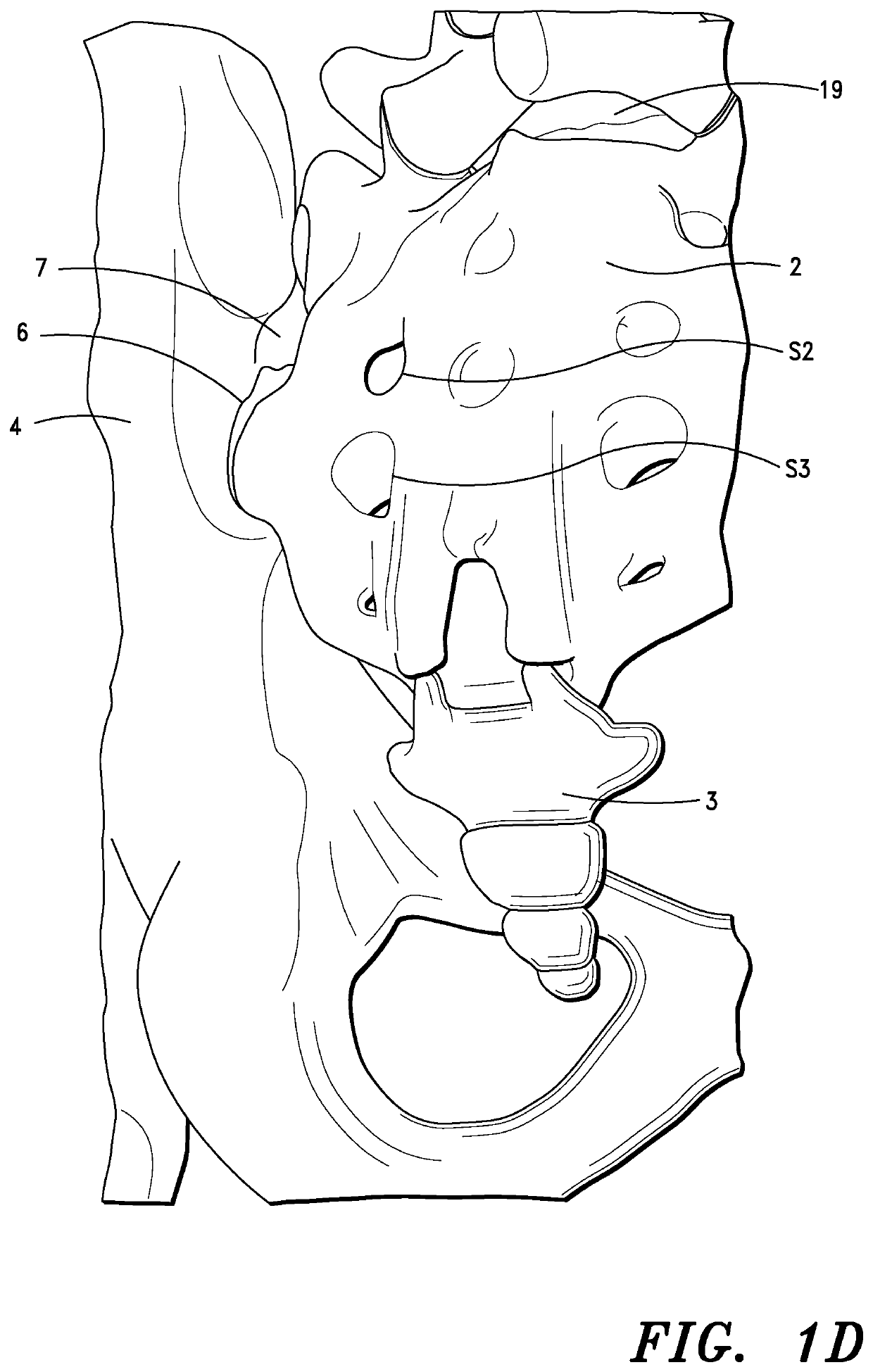
Further, some conventional minimally-invasive SI joint stabilization methods are particularly prone to failure due to displacement of the SI joint prostheses in the dysfunctional SI joint, such as in or 
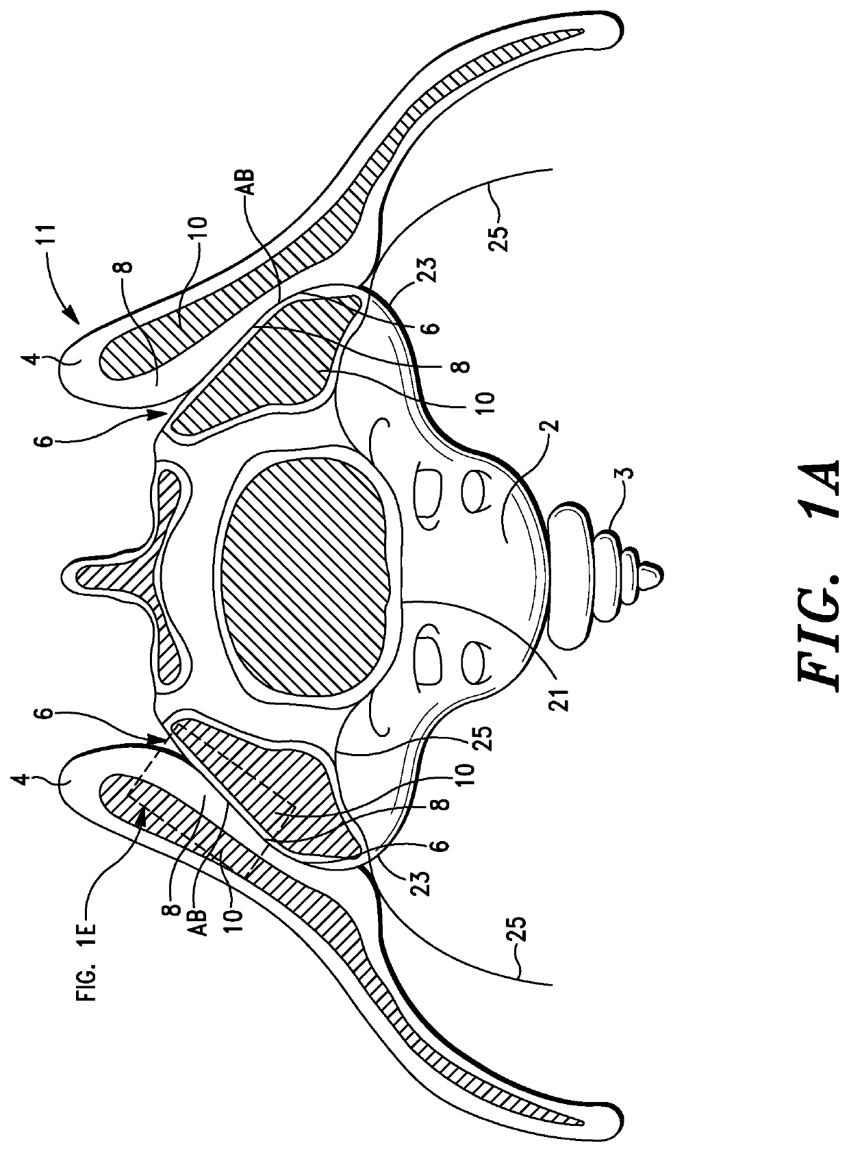
proximate the SI joint dorsal recess referenced above and shown in FIG. 1D, and / or failure of the prostheses to effectively engage the SI joint structures, e.g., articular surfaces of the sacrum and / or ilium.
Although many of the “improved” SI joint prostheses, when deployed properly in a dysfunctional SI joint, can, and often will, effectively engage SI joint structures, there remains several disadvantages associated with the prostheses.
A major disadvantage associated with the noted SI joint prostheses is that the liquefiable synthetic polymers, when re-solidified in situ, are structurally inferior to the osseous or bone tissue of the sacrum and ilium.
The fusion sites between the articular surfaces of the sacrum and ilium that define the SI joint are, thus, highly susceptible to structural fatigue and failure, which can, and often will, result in misalignment of the SI joint and ultimately increased pain for the subject.
A further disadvantage associated with the SI joint prostheses disclosed in U.S. Pat. No. 8,951,254 is that the synthetic liquefiable synthetic polymers are also substantially immunogenic and will induce an adverse immune response when the prostheses are implanted in a dysfunctional SI joint.

Method used


Image

  • Methods for Sacroiliac Joint Stabilization
  • Methods for Sacroiliac Joint Stabilization
  • Methods for Sacroiliac Joint Stabilization

Examples


examples

[0539]The following example is provided to enable those skilled in the art to more clearly understand and practice the present invention. The example should not be considered as limiting the scope of the invention, but merely as being illustrated as representative thereof.

[0540]An adult male, age 42 presented with a traumatic injury proximate the SI joint, resulting in a dysfunctional SI joint and significant pain associated therewith, i.e., a visual analog pain score (VAS) of approximately 8.0.

[0541]CT scans were initially performed to determine the full extent of the patient's injury, check for any SI joint abnormalities, and plan the SI joint stabilization procedure, including determining the incision site, guide pin trajectory, and SI joint prosthesis required to stabilize the dysfunctional SI joint.

[0542]Before proceeding with the SI joint stabilization procedure, CT scans were also performed to ensure proper alignment of the patient on the surgical table.

[0543]The SI joint sta...


PUM


Abstract

Methods are described for conducting minimally invasive medical interventions utilizing instruments and assemblies thereof to stabilize and / or fixate a dysfunctional sacroiliac (SI) joint. The methods include the initial steps of providing a drill guide assembly adapted to create a pilot opening in the dysfunctional SI joint, a prosthesis configured to be inserted into the pilot opening created by the drill guide assembly and a prosthesis deployment assembly adapted to engage and advance the prosthesis into the pilot opening in the dysfunctional SI joint. The drill guide assembly includes a drill guide having a prosthesis access opening therethrough; the opening having a configuration that corresponds to the shape of the prosthesis. In some aspects of the invention, the methods thus include the step of advancing the prosthesis through the drill guide and then into the pilot opening in the dysfunctional SI joint with the prosthesis deployment assembly; the initial advancement of the prosthesis through the drill guide providing consistent, optimal placement of the prosthesis in the dysfunctional SI joint.

Description

CROSS-REFERENCES TO RELATED APPLICATIONS[0001]This application is a continuation-in-part of U.S. application Ser. No. 17 / 833,098, filed Jun. 6, 2022, which is a continuation of U.S. patent application Ser. No. 17 / 749,199, filed on May 20, 2022, which is a continuation-in-part application of U.S. patent application Ser. No. 17 / 740,568, filed on May 10, 2022, which is a continuation-in-part application of U.S. patent application Ser. No. 17 / 463,779, filed Sep. 1, 2021, which is a continuation-in part of U.S. patent application Ser. No. 13 / 857,977, filed Apr. 5, 2013, now U.S. Pat. No. 11,273,042, which is a continuation application of U.S. patent application Ser. No. 13 / 192,289, filed Jul. 27, 2011, now abandoned, which claims the benefit of U.S. provisional patent application Ser. No. 61 / 368,233, filed Jul. 27, 2010.FIELD OF THE INVENTION[0002]The present invention relates to methods, systems and apparatus for stabilizing junctions between bone structures. More particularly, the pres...

Claims


Application Information

Patent Timeline
IPC IPC(8): A61F2/30A61B17/70A61B34/20A61B17/88A61B17/16A61B6/00A61B6/03A61B8/08A61B5/055A61F2/44A61F2/46A61B5/06
CPCA61F2/30988A61B17/70A61B34/20A61B17/8858A61B17/1604A61B6/485A61B6/032A61B17/7074A61B8/0841A61B5/055A61B17/7055A61B17/1664A61F2/4455A61B17/1671A61F2/4601A61B5/062A61F2/4611A61F2002/30995A61F2002/30121A61F2002/30166A61F2002/30123A61F2002/30593A61B2576/00A61B5/0036A61B5/4528A61B5/4571A61B8/0875A61B17/1757A61B2017/0046A61B2090/374A61B2090/376A61B2090/3762A61B2090/378A61B2505/05A61F2/30767A61F2/447A61F2/4603A61F2002/30772A61F2002/30777A61F2002/30784A61F2002/30845A61F2002/3093A61F2002/4625A61F2002/4627A61F2002/4629A61F2002/4631A61F2002/4635A61F2002/4677A61F2002/4681A61B6/508A61B6/505A61B6/487A61B6/12A61B17/848A61B2090/062A61B2090/3966A61F2002/30064A61F2002/3013A61F2002/30622A61F2002/3068A61F2002/3085
Inventor GINN, RICHARD S.BROWN, RICHARDSTEWART, DAREN
Owner TENON MEDICAL INC