Method and system for pre-determining spectral doppler user parameters

a user parameter and spectral doppler technology, applied in the field of ultrasonic imaging techniques, can solve the problems of cumbersome manual angle correction of blood flow velocity, time-consuming examination process, and difficult repeatability

Inactive Publication Date: 2001-01-23
SIEMENS MEDICAL SOLUTIONS USA INC
View PDF7 Cites 62 Cited by
  • Summary
  • Abstract
  • Description
  • Claims
  • Application Information

AI Technical Summary

Problems solved by technology

As noted in the Banjanin et al. patent, the manual angle correction of blood flow velocity is often cumbersome and hard to repeat.
Since there are a number of operation parameters that must be set when the spectral Doppler mode is initiated, the examination process is often time consuming.
While this basis for establishing initial settings reduces the time required for an examination, there are a large number of variables that play a role in determining the settings, so that very few examinations match the typical examination.

Method used


Image

  • Method and system for pre-determining spectral doppler user parameters
  • Method and system for pre-determining spectral doppler user parameters
  • Method and system for pre-determining spectral doppler user parameters

Examples


Embodiment Construction

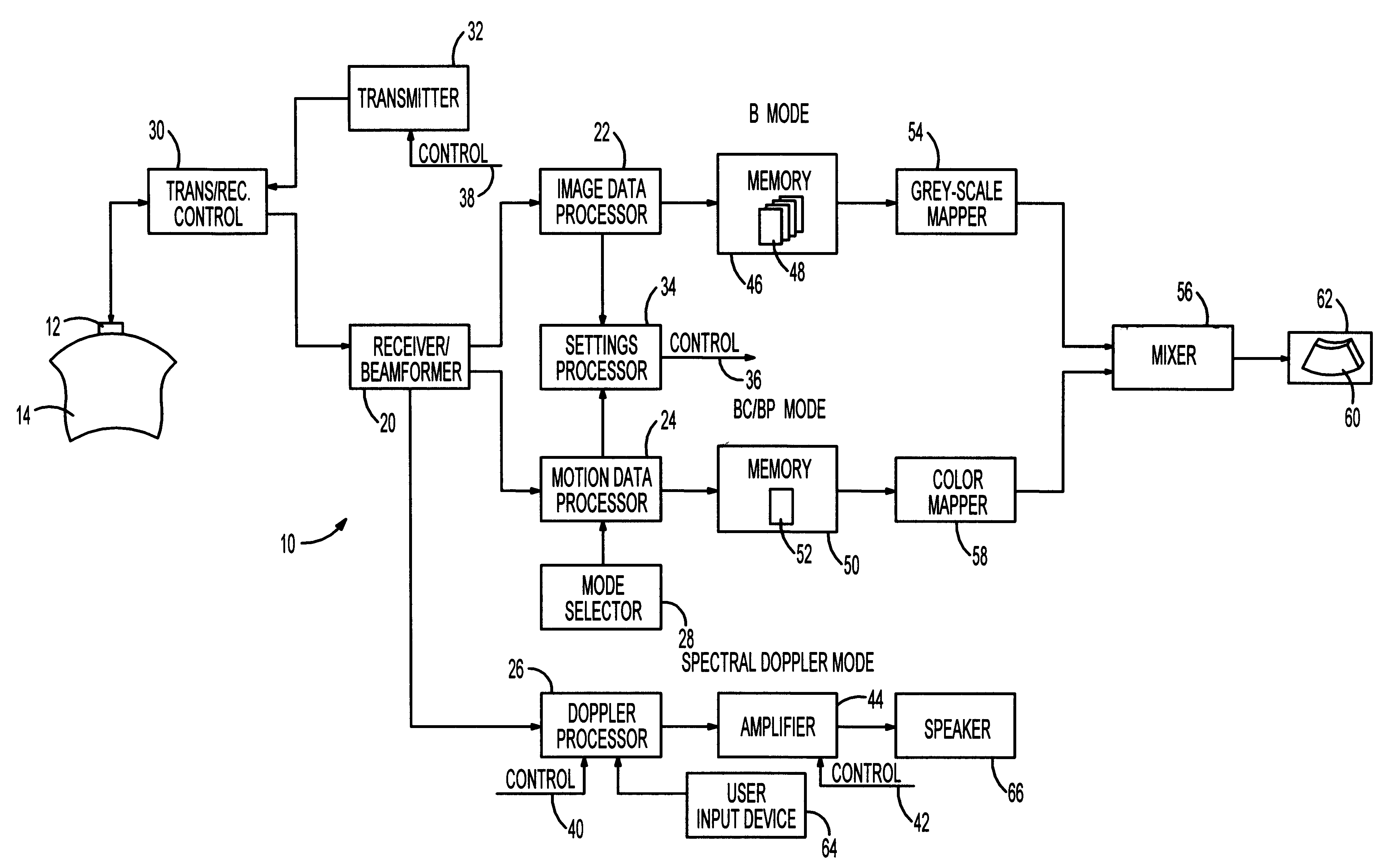
The process flow of FIG. 4 is utilized when the ultrasonic system 10 of FIG. 1 is switched into the spectral Doppler mode from B mode operation. Typically, B mode imaging is executed simultaneously with colorflow or power imaging. Since the colorflow and power mode operations provide more information than is generated by B mode imaging, the process of FIG. 4 is followed only when the system is limited to B mode imaging.

Steps 122, 124 and 126 are conventional steps of initiating the B mode operation, acquiring ultrasound-based data, and processing the acquired data to generate image information. In FIG. 1, the transducer generates ultrasonic beams and forms electrical signals that are responsive to echoes of the beam energy from the patient 14. This acquired ultrasound-based data is processed at the image data processor 22. In step 128, the memory 46 stores frames 48 of the image information. The grey-scale mapper 54 is utilized to generate images at the display unit 62. In FIG. 4, t...


PUM


Abstract

A method of initializing a spectral Doppler mode of operation for an ultrasonic system includes acquiring ultrasound-based data during a two-dimensional mode of operation for a particular examination session and includes automatically establishing settings for the Doppler mode operation parameters based upon the ultrasound-based data. That is, the ultrasound-based data is processed during the session to select spectral Doppler mode settings that are specific to the ongoing session. The session-specific settings are invoked when the system is switched to the spectral Doppler mode. If the two-dimensional mode is a colorflow mode, the Doppler sample volume can be based upon detecting the location of maximum velocity in the colorflow image, the angle correct setting can be based upon maximum velocities in colorflow vectors, the pulse repetition frequency setting can be based upon the maximum frequency shift detected in the colorflow data, and the gain setting can be based upon the amplitude of colorflow data. On the other hand, if the two-dimensional mode is a power mode, the Doppler sample volume is based upon detecting the region of the power mode image having the strongest signals, the angle correct setting can be based upon detecting the direction of flow, the Doppler pulse repetition frequency can be based upon a scale setting for the power mode data acquisition, and a gain setting can be based upon the amplitude of power mode data. Lastly, if the two-dimensional mode is the grey-scale imaging mode, the selection of a Doppler sample volume can be based upon identifying a dark region near the center of the grey-scale image, the angle correct setting can be based upon the orientation of the boundaries of the identified dark region, and the gain setting can be based upon the amplitude of image data.

Description

The invention relates generally to a method and a system for selecting settings for process parameters in an ultrasonic examination and relates more particularly to selecting settings for process parameters upon switching an ultrasonic system from a two-dimensional mode of operation to a spectral Doppler mode of operation.DESCRIPTION OF THE RELATED ARTUltrasonic imaging techniques may be used to produce images of internal features of an object, such as tissues of a human body. A diagnostic ultrasonic imaging system for medical use forms images of internal tissues of the human body by electrically exciting an acoustic transducer element or an array of acoustic transducer elements to generate ultrasonic beams that propagate into the patient's body. Energy of the ultrasonic beams reflects off human tissues that present discontinuities or impedance changes to the propagating ultrasonic beams. The echoes from the tissues are sensed by the transducer and are converted into electrical sign...

Claims


Application Information

Patent Timeline
Patent Type & Authority Patents(United States)
IPC IPC(8): A61B8/06G01S15/89G01S15/00G01S7/52G01S15/58
CPCA61B8/06A61B8/13G01S7/5206G01S7/52066G01S7/52071G01S15/8979G01S15/8984G01S15/8988
Inventor FREIBURGER, PAUL D.
Owner SIEMENS MEDICAL SOLUTIONS USA INC
Why Eureka
Try Eureka