Implantable monitor

a monitor and monitor technology, applied in the field of implantable monitors, can solve the problems of power consumption, externally worn ecg recorders have inherent limitations in the memory capacity of storing sampled ecg and egm data, and the inability to induce and observe palpitation symptoms by the physician

Inactive Publication Date: 2001-05-08
MEDTRONIC INC
View PDF11 Cites 681 Cited by
  • Summary
  • Abstract
  • Description
  • Claims
  • Application Information

AI Technical Summary

Problems solved by technology

Frequently faintness, syncope, and tachyarrhythmia palpitation symptoms cannot be induced and observed by the physician in tests conducted in a clinic.
As described in commonly assigned U.S. Pat. No. 5,312,446 and in U.S. Pat. No. 4,947,858, both incorporated herein by reference, the externally worn ECG recorders have inherent limitations in the memory capacity for storing sampled ECG and EGM data.
Cost, size, power consumption, and the sheer volume of data over time have limited real time external Holter monitors to recording 24 hour segments or recording shorter segments associated with arrhythmias that are felt by the patient who initiates storage.
The use of the externally worn Holter monitor coupled with skin electrodes is also inconvenient and uncomfortable to the patient.
The skin electrodes can work loose over time and with movement by the patient, and the loose electrodes generates electrical noise that is recorded with the EGM signal and makes its subsequent analysis difficult.
However, many times patients are either unaware of "silent" cardiac arrhythmias or are asleep or fail to activate the recording function when they recover from syncope (i.e., have fainted) when bradycardias and tachyarrhythmias occur, and so the accompanying EGM episode data is not recorded.
But, the subcutaneous location environment of the sense electrode pair or pairs on the device housing is relatively noisy due to electromyographic signals generated by adjacent muscle groups that are exercised by the patient.
On the other hand, sometimes such noise is present during an actual arrhythmia of interest that is correctly detected, triggering the recording of EGM episode data that is of interest but is also noisy.
The physician may find that the EGM episode data that is later uplink transmitted and displayed by the programmer is simply corrupted and of no value in diagnosing the patient's cardiac condition.

Method used


Image

  • Implantable monitor
  • Implantable monitor
  • Implantable monitor

Examples


Embodiment Construction

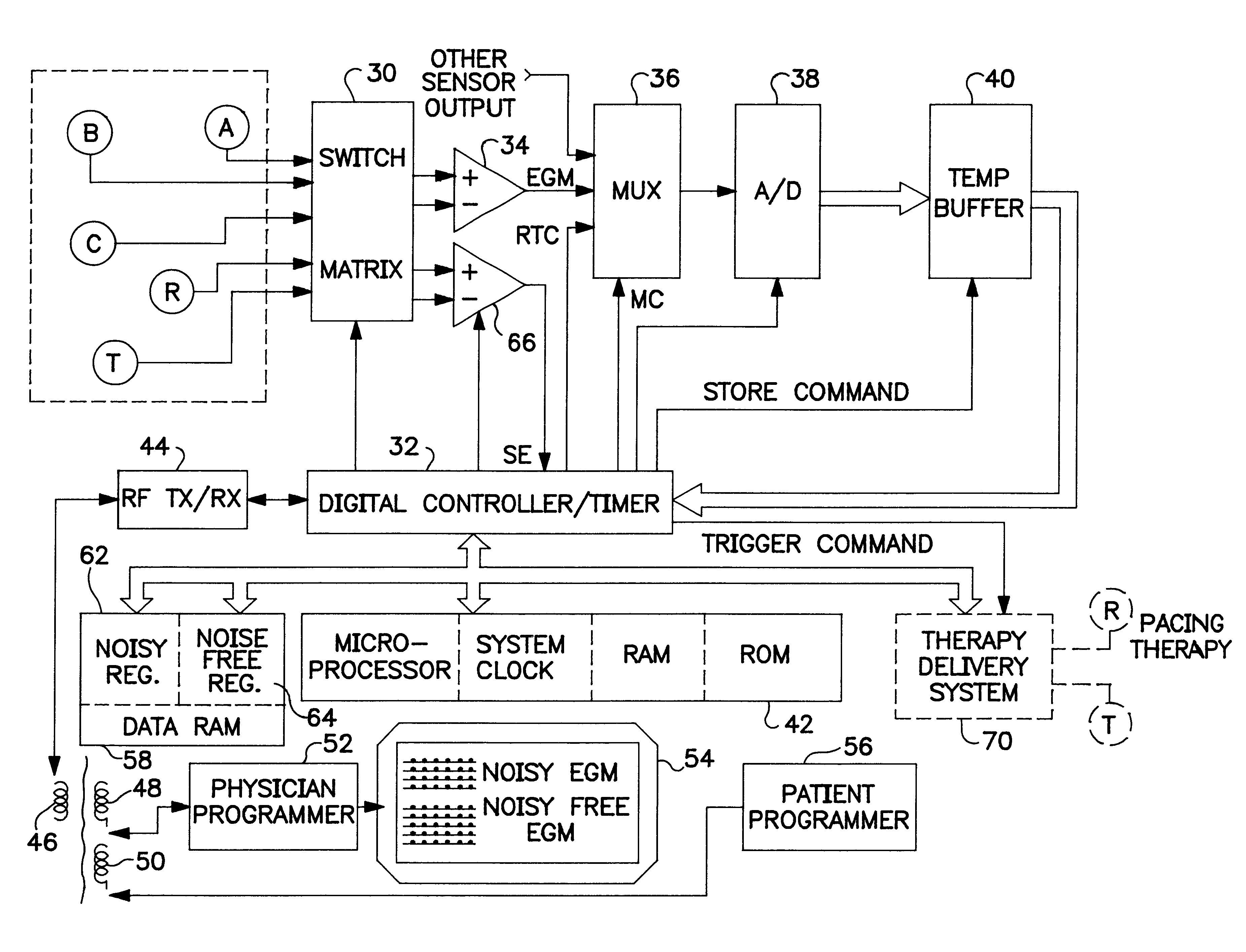
The noise discrimination and data storage methods and apparatus of the present invention find particular utility in cardiac EGM monitors that have EGM sense electrodes locate where electromyographic noise can contaminate the EGM signal and corrupt it. Consequently, they will be described in conjunction with such monitors or therapy delivery IMDs as illustrated in FIGS. 1 and 2. However, it will be understood that the present invention is not so limited and that the noise discrimination and data storage methods and apparatus of the present invention can be used in other cardiac or other electrical signal monitors implanted in the body or coupled externally to the body.

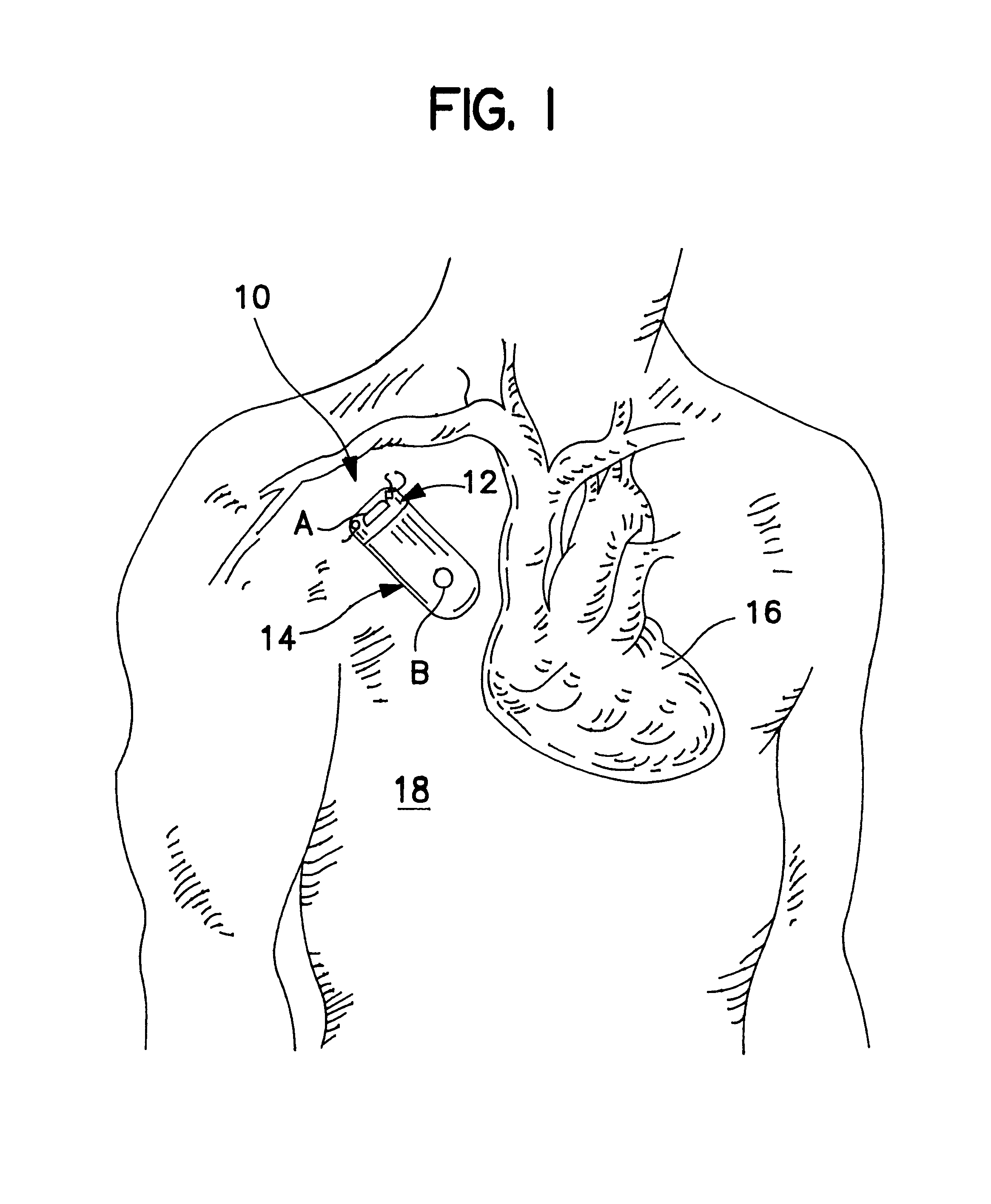
FIG. 1 is a simplified schematic view of an implantable cardiac monitor 10 embodying the improvements of the present invention implanted subcutaneously in the upper thoracic region of the patient's body 18 and displaced from the patient's heart 16. The housing of the cardiac monitor 10 takes the shape of the MEDTRONIC.R...


PUM


Abstract

An implantable medical device (IMD) capable of monitoring physiologic data, distinguishing relatively noisy and noise free physiologic data, and recording noisy and relatively noise free segments of physiologic data in separate memory registers of a limited memory for retrieval and analysis at a later time. Preferably the physiologic data comprises the sampled EGM of the heart detected from sense electrode pairs that are implanted in the patient at sites where extraneous electrical noise, e.g., electromyographic signals, are also capable of being detected. The sense electrode pairs can constitute one or both sense electrodes located on or adjacent to the atrial and / or ventricular heart chambers and coupled to the IMD by a lead body or sense electrode pairs that are located remotely from the heart, e.g. at a subcutaneous implantation site of the IMD. A plurality of noisy EGM episode data registers store a corresponding plurality of noisy EGM episode data sets on a FIFO basis and another plurality of noise free EGM episode data registers to store a corresponding plurality of relatively noise free EGM episode data sets on a FIFO basis. Any form of discrimination of noisy data from relatively noise free data can be employed at the time of recording, but because the stored EGM episode data sets are subsequently viewed and analyzed by a physician, discrimination with absolute certainty is not required, and the physician can alter the detection criteria to fine tune it.

Description

The present invention relates to discrimination of noisy physiologic data from relatively noise free physiologic data and selective storage thereof in an implantable medical device (IMD) and more particularly to methods and apparatus for discriminating noisy and relatively noise-free cardiac electrogram (EGM) episode data and selectively storing the noisy and noise-free EGM episode data in separate EGM data registers for later retrieval and analysis by a physician.There are many instances where it is desirable to be able to diagnose intermittent spontaneous cardiac arrhythmias in ambulatory patients. Frequently faintness, syncope, and tachyarrhythmia palpitation symptoms cannot be induced and observed by the physician in tests conducted in a clinic. For many years, such patients have been equipped with external ECG monitoring systems, e.g., the patient-worn, real time Holter monitors, that continuously sample the ECG from skin electrodes and record it over a certain time period. The...

Claims


Application Information

Patent Timeline
Patent Type & Authority Patents(United States)
IPC IPC(8): A61B5/00A61N1/37A61N1/362
CPCA61B5/0008A61B5/0031A61N1/3702A61B5/7203A61B5/7207
Inventor DUFFIN, EDWIN G.
Owner MEDTRONIC INC
Why Eureka
Try Eureka