
Radiation breast cup and method

a breast cup and breast cup technology, applied in the field of breast cup and method, can solve the problems of advanced adverse conditions, extremely uncomfortable plates and procedures for patients, etc., and achieve the effect of less painful and uncomfortabl

Inactive Publication Date: 2002-07-09
BROADNAX JUANITA L
View PDF7 Cites 28 Cited by
  • Summary
  • Abstract
  • Description
  • Claims
  • Application Information

AI Technical Summary

Benefits of technology

X-ray imaging and other types of radiation therapy have become increasingly common in recent years. Tumors and other tissue changes which may become cancerous are often detected through images made by such techniques. Mammography is a popular procedure which requires the breast tissues to be compressed, usually between one or more plates during radiation. While such compression is necessary for a better image, such plates and procedures are extremely uncomfortable for the patient and prevent, or at least discourage, such examinations for many. Sometimes delays in obtaining radiation examinations cause greatly advanced adverse conditions which may be detected too late. Thus, as breast tissues require compression for well-defined images, it is an objective of the present invention to provide a method of compressing the breast tissue which is less painful and uncomfortable than conventional techniques.
It is another objective of the present invention to provide a stretchable breast cup which can be easily fitted to allow even pressure for a clear, readable and usable x-ray.
It is still another objective of the present invention to provide a breast cup which can be easily worn with little training.

Problems solved by technology

While such compression is necessary for a better image, such plates and procedures are extremely uncomfortable for the patient and prevent, or at least discourage, such examinations for many.
Sometimes delays in obtaining radiation examinations cause greatly advanced adverse conditions which may be detected too late.

Method used


Image

  • Radiation breast cup and method
  • Radiation breast cup and method
  • Radiation breast cup and method

Examples


Embodiment Construction

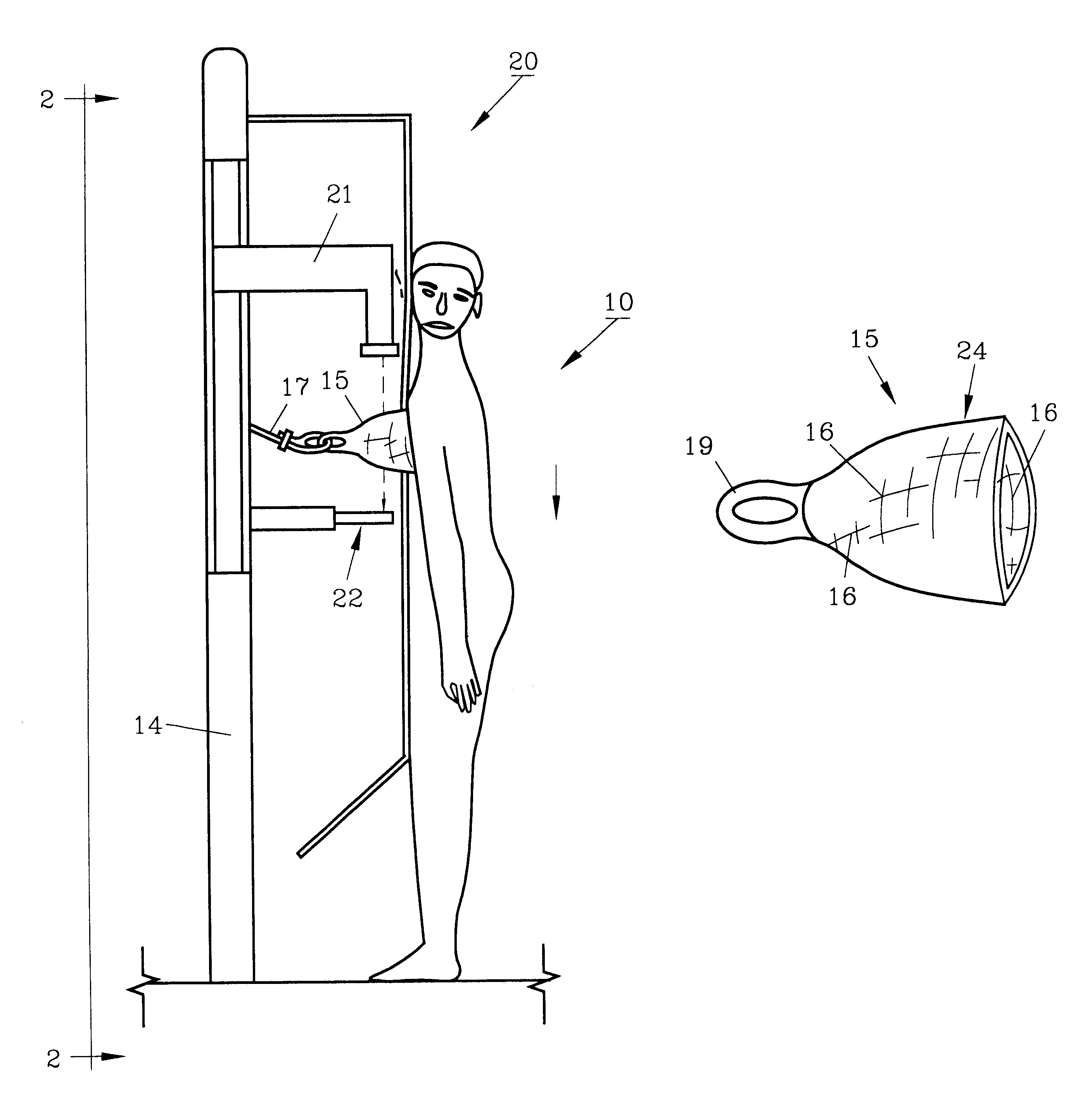
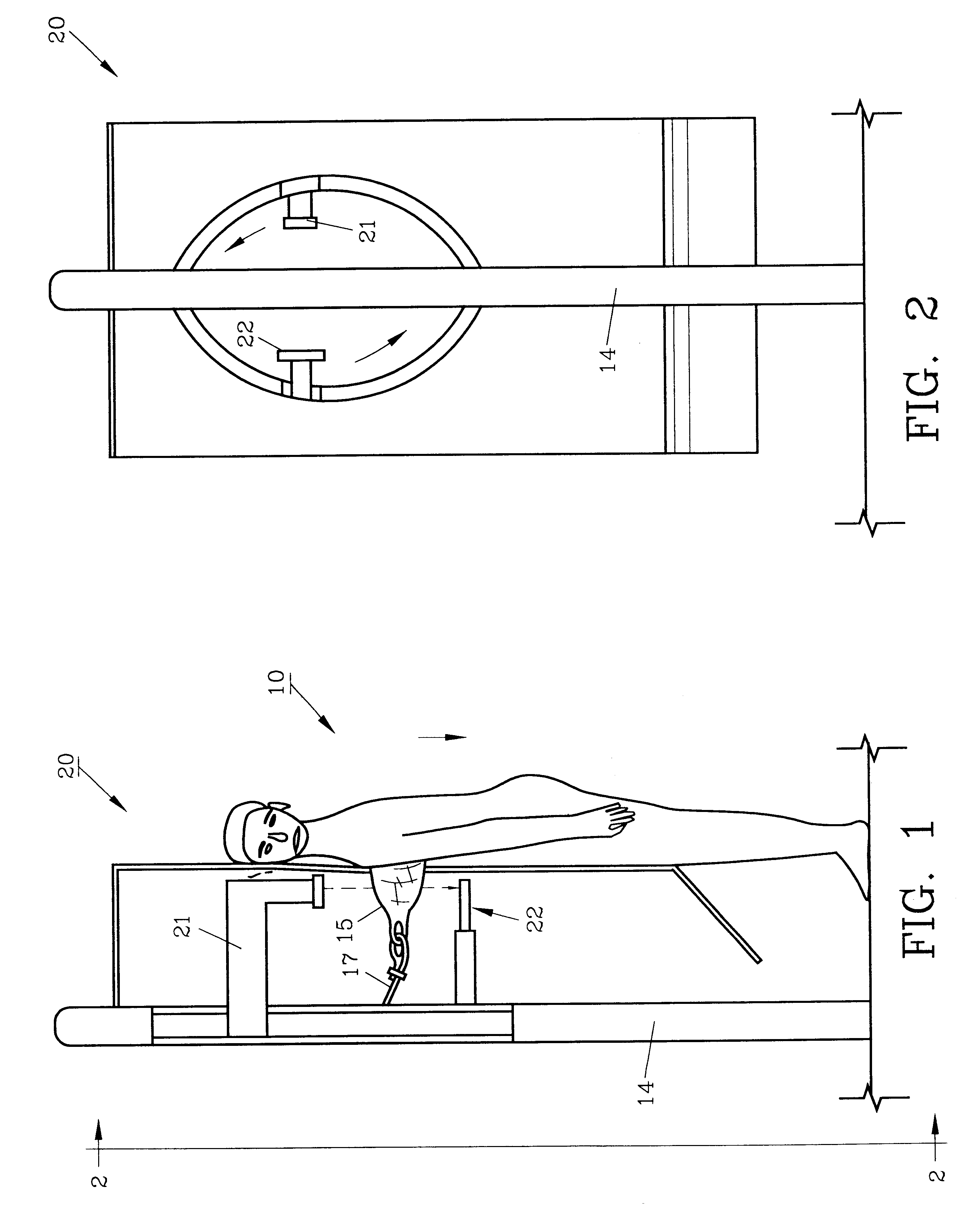
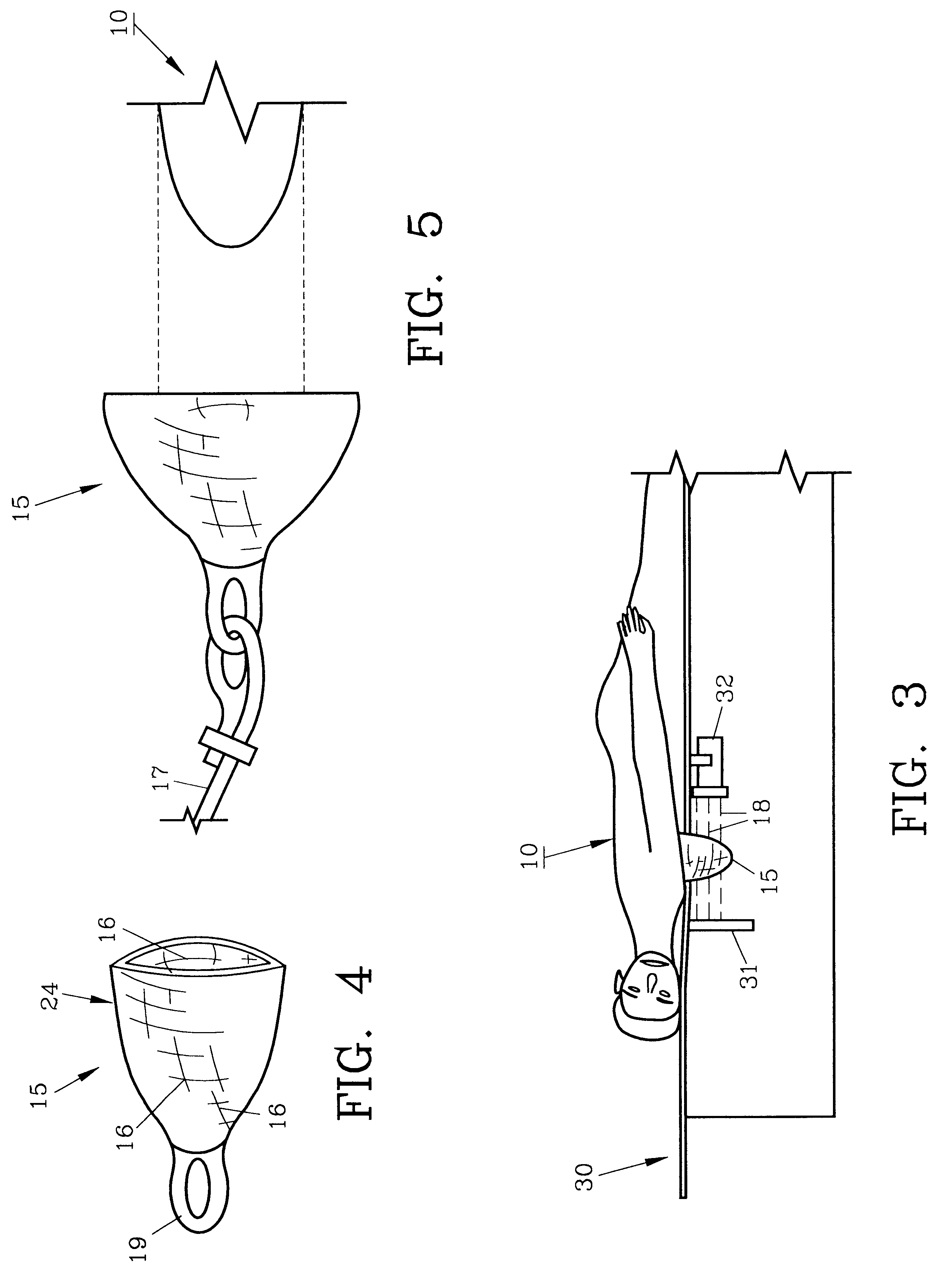
For a better understanding of the invention and its operation, turning now to the drawings, FIG. 1 demonstrates female patient 10 in a standing position against conventional x-ray system 20 as used in mammography. Breast cup 15 is worn by female patient 10 and it is joined to x-ray system stanchion 14 by flexible cord 17. Eyelet 19 joins breast cup 15 to cord 17 as seen in FIG. 4. As would be understood, radiation 18 passes from generator 21 through breast cup 15 to image receiver 22 which may for example, contain x-ray film, lenses or the like. Image receiver 22 is contiguous breast cup 15 as viewed in 1.

X-ray system 20 in FIG. 2 allows x-ray generator 21 and image receiver 22 to each revolve in a somewhat circular pattern to selected locations for imaging at various angles as desired, (see also FIG. 7). FIG. 3 demonstrates female patient 10 in a prone posture on x-ray system 30 wearing breast cup 15 proximate x-ray generator 32 and image receiver 31, seen in schematic representati...


PUM


Abstract

A breast cup is described herein which is made from a stretchable material for compressing the breast during x-ray imaging. The breast cup is constructed preferably of a fabric having elastomeric yarns to allow it to be easily worn during radiation and afterwards removed with minimal effort. The stretchable qualities provide the needed compression throughout the cup, yet is not unduly uncomfortable while worn.

Description

The invention herein pertains to radiation imaging procedures as are used in mammography and specifically pertains to a breast cup which can be used to x-ray the breast without undue discomfort and can be easily expanded / contracted to fit various breast sizes.DESCRIPTION OF THE PRIOR ART AND OBJECTIVES OF THE INVENTIONX-ray imaging and other types of radiation therapy have become increasingly common in recent years. Tumors and other tissue changes which may become cancerous are often detected through images made by such techniques. Mammography is a popular procedure which requires the breast tissues to be compressed, usually between one or more plates during radiation. While such compression is necessary for a better image, such plates and procedures are extremely uncomfortable for the patient and prevent, or at least discourage, such examinations for many. Sometimes delays in obtaining radiation examinations cause greatly advanced adverse conditions which may be detected too late. ...

Claims


Application Information

Patent Timeline
Patent Type & Authority Patents(United States)
IPC IPC(8): A61B6/04
CPCA61B6/0414A61B6/502
Inventor BROADNAX, JUANITA L.
Owner BROADNAX JUANITA L
Learn More