Bone marrow cells as a source of neurons for brain and spinal cord repair

a bone marrow cell and brain technology, applied in the field of bone marrow cells as a source of neurons for brain and spinal cord repair, can solve the problems of no effect of these therapeutic measures on the underlying disease process, no longer being able to compensate for the loss of basal ganglia dopamine, and difficult if not impossible to rebuild

Inactive Publication Date: 2003-03-04
SOUTH FLORIDA UNIVESITY OF
View PDF14 Cites 196 Cited by
  • Summary
  • Abstract
  • Description
  • Claims
  • Application Information

AI Technical Summary

Problems solved by technology

Neurobiologists have long considered the neurons in the adult brain to be like a precious nest egg: a legacy that dwindles with time and illness and is difficult if not impossible to rebuild.
Although the modem treatment of PD is more successful than any that was available before the introduction of levodopa, including stereotactic surgery, there are still many problems.
Underlying much of the difficulty undoubtedly is the fact that none of these therapeutic measures has an effect on the underlying disease process, which consists of neuronal degeneration.
Ultimately, a point seems to be reached where pharmacology can no longer compensate for the loss of basal ganglia dopamine.
At present there are palliative treatments but no means to restore function.
Purkinje cells are permanently lost, resulting in ataxia.
Even if the patient is permanently cured of the cancer, their ability to function may be profoundly disabled by the loss of Purkinje cells.
Strokes also result in neuronal degeneration and loss of functional synapses.
The experimental replacement of neurons by direct grafting of fetal tissue into the brain has been accomplished in small numbers of patients in several research universities (including our University of South Florida); but so far, the experimental grafting of human fetal neurons has been limited by scarcity of appropriate tissue sources, logistic problems, legal and ethical constraints, and poor survival of grafted neurons in the human host brain.
Even though the multipotential properties of MSCs have been recognized for several decades, there are surprisingly large gaps in our information about the cells themselves.
The isolation of adequate numbers of HSCs requires large volumes of marrow (1 liter or more), and the cells are difficult to expand in culture.

Method used


Image

  • Bone marrow cells as a source of neurons for brain and spinal cord repair

Examples


example 1

In Vitro Differentiation

Bone marrow was obtained from either mouse femurs or from human bone marrow aspirates. Human bone marrow was diluted 1:1 with Dulbecco's Minimal Essential Media (DMEM), (GIBCO / BRL) and 10% fetal bovine serum (FBS) and centrifuged through a density gradient (Ficoll-Paque Plus, 1.077 g / ml, Pharmacia) for 30 min at 1,000 g. The supernatant and interface were combined, diluted to approximately 20 ml with MEM with 10% fetal calf serum (FCS) and plated in polyethylene-imine coated plastic flasks. Mouse bone marrow was placed in 10 ml PBS and 5% bovine albumin. Cells were washed in this medium and centrifuged at 2000 rpm for 5 min. Cells were resuspended in growth medium consisting of DMEM supplemented with 2mM glutamine, 0.001% .beta.-mercaptoethanol, non-essential amino acids and 10% horse serum. The cells were incubated in the flasks for two days after which non-adherent cells were removed by replacing the medium. After the cultures reached confluency, the cells ...

example 2

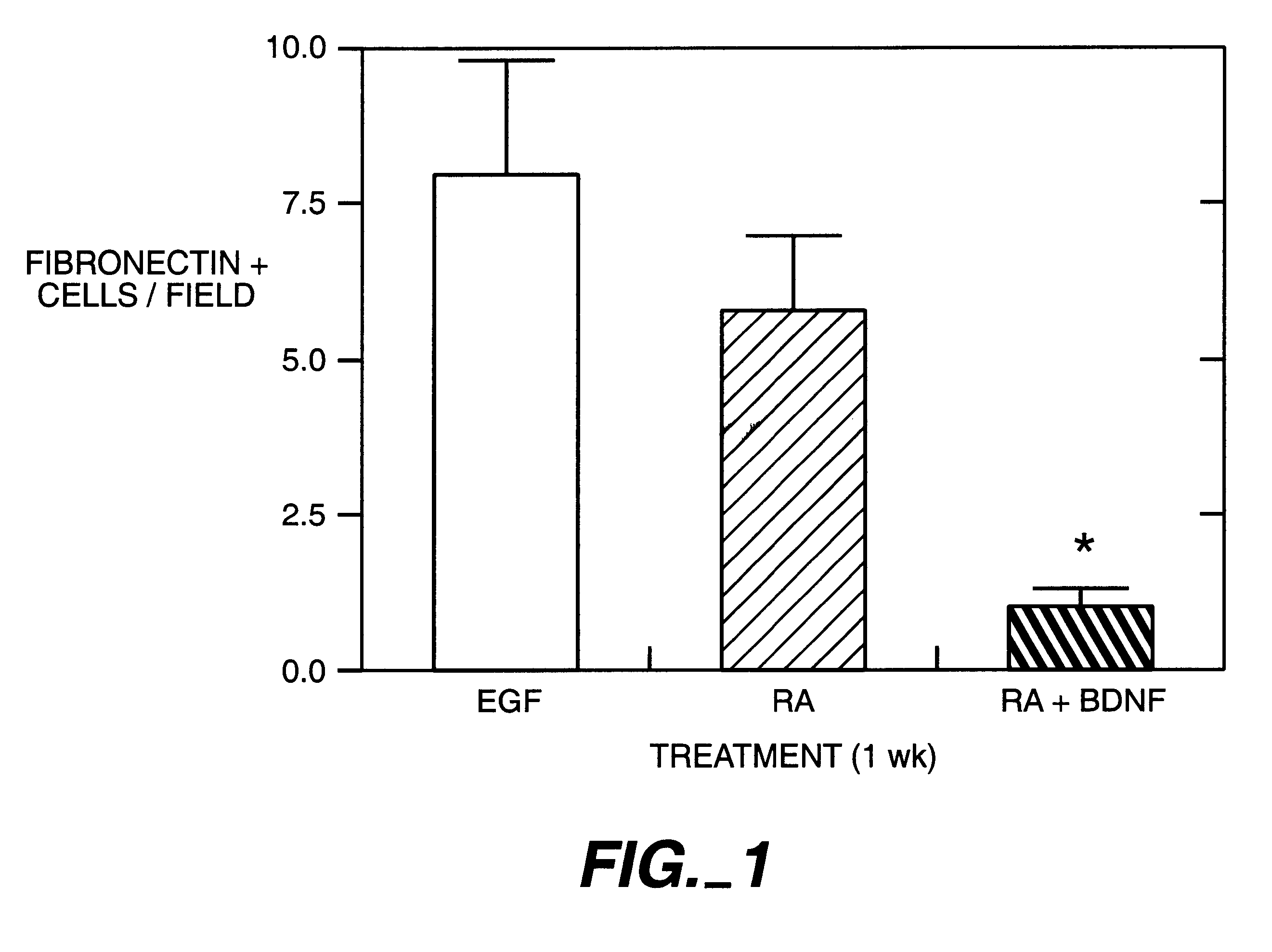
Bone marrow cells were induced to become neuron-like cells in vitro employing methods similar to those used to promote differentiation of embryonic stem (ES) cells. J. Dinsmore, et al., Cell Transplantation 5:131-143 (1996). Bone marrow was obtained from adult mice and divided into two fractions using magnetic cell sorting. First, bone marrow cells were collected from mouse femur and tibias by flushing the shaft with buffer (PBS supplemented with 0.5% BSA, pH 7.2) using a syringe with a #26 G needle. Cells were disaggregated by gentle pipetting several times. Cells were passed through 30 .mu.m nylon mesh to remove remaining clumps of tissue. Cells were washed by adding buffer, centrifuging for 10 min at 200Xg and removing supernatant. The cell pellet was resuspended in 800 .mu.L of buffer for each 10.sup.8 cells. With a magnetic cell sorting kit (Milteny Biotec, Inc, Auburn Tex.), hematopoietic bone marrow cells were labeled with Scal+microbeads. The labeled bone marrow cells were p...

example 3

In Vivo Differentiation

To determine whether differentiation of BMSC cells into neurons would occur in vivo, BMSC from lacZ mice were grafted into denervated or normal rat striatum. Prior to grafting, BMSC were treated for 2 days with cis-9 retinoic acid (0.5 .mu.M) and BDNF (10 ng / ml). Three weeks prior to grafting, the graft site of the caudate / putamen of rats was treated with unilateral intranigral injections of 6-hydroxydopamine (6-OH DA). A single injection of 6-OH DA (Sigma; 2.5 .mu.L, 3.6 .mu.g / .mu.L for a total dose of 9 .mu.g in 0.2% ascorbic acid) was made into the right ascending mesostriatal dopaminergic system (4.4 mm posterior to bregma, -1.2 mm laterally and -7.8 mm ventral to dura with the toothbar set at -2.4 mm). The 6-OH DA was delivered at a rate of 1 .mu.L / min. The syringes were held in place for an additional 5 min before slow withdrawal.

BMSC suspension aliquots were deposited in the striatum along a single needle tract on the same side as the 6-OH DA nigral les...


PUM

PropertyMeasurementUnit
timeaaaaaaaaaa
volumesaaaaaaaaaa
densityaaaaaaaaaa

Abstract

Bone marrow stromal cells (BMSC) differentiate into neuron-like phenotypes in vitro and in vivo, engrafted into normal or denervated rat striatum. The BMSC did not remain localized to the site of the graft, but migrated throughout the brain and integrated into specific brain regions in various architectonic patterns. The most orderly integration of BMSC was in the laminar distribution of cerebellar Purkinje cells, where the BMSC-derived cells took on the Purkinje phenotype. The BMSC exhibited site-dependent differentiation and expressed several neuronal markers including neuron-specific nuclear protein, tyrosine hydroxylase and calbindin. BMSC can be used to target specific brain nuclei in strategies of neural repair and gene therapy.

Description

1. Field of UseThis application relates to methods of culturing bone marrow cells such that they express neuronal phenotype for use in transplantation.2. Background InformationNeurobiologists have long considered the neurons in the adult brain to be like a precious nest egg: a legacy that dwindles with time and illness and is difficult if not impossible to rebuild. Parkinson's and Alzheimer's are examples of neurodegenerative diseases whose cures await scientists overcoming the difficulty of rebuilding neurons in the human adult brain. Parkinson's disease (PD), is a disorder of middle or late life, with very gradual progression and a prolonged course. HARRISON'S PRINCIPLES OF INTERNAL MEDICINE, Vol. 2, 23d ed., Ed. by Isselbacher, Braunwald, Wilson, Martin, Fauci and Kasper, McGraw-Hill Inc., New York City, 1994, pg. 2275. The most regularly observed changes have been in the aggregates of melanin-containing nerve cells in the brainstem (substantia nigra, locus coeruleus), where ther...

Claims


Application Information

Patent Timeline
Patent Type & Authority Patents(United States)
IPC IPC(8): C12N5/06A61K35/12C12N15/09A61K35/28A61K49/00A61P43/00C12N5/02C12N5/0793C12N5/10
CPCC12N5/0619A61K35/12A61K2035/124C12N2500/46C12N2501/11C12N2506/1353C12N2501/13C12N2501/385C12N2501/392C12N2502/08C12N2501/115A61P43/00
Inventor SANCHEZ-RAMOS, JUANSONG, SHIJIEJANSSEN, WILLIAMSANBERG, PAULFREEMAN, THOMAS
Owner SOUTH FLORIDA UNIVESITY OF
Why Eureka
Try Eureka