Hematopoietic growth factor inducible neurokinin-1 gene and uses thereof

a technology of hematopoietic growth factor and neurokinin, which is applied in the direction of genetic material ingredients, peptides, drug compositions, etc., can solve the problems of stem cells, bone marrow may not produce enough stem cells, breast cancer metastasis to the bone marrow, etc., to reduce the toxicity of normal tissues and increase the effectiveness of chemo- and radiotherapy

Inactive Publication Date: 2008-03-13
RUTGERS THE STATE UNIV
View PDF1 Cites 13 Cited by
  • Summary
  • Abstract
  • Description
  • Claims
  • Application Information

AI Technical Summary

Benefits of technology

[0030] Hence, the methods of the present invention are useful for the prevention and treatment of lymphoproliferative syndromes such as B-cell related maladies, including but not limited to acute and chronic myeloid and lymphocytic leukemia as well as the B-cell subtype of Hodgkin's and non-Hodgkin's lymphomas. Further, the methods of the present invention can be used to increase the effectiveness of both chemo- and radiotherapy. Further still, the use of monoclonal antibodies, in conjunction with the gene, antisense polynucleotide or protein of the present invention, to direct radionuclides, toxins, or other therapeutic agents offers the possibility that such agents can be delivered at lower dosages, selectively to tumor sites, thus limiting toxicity to normal tissues.

Problems solved by technology

Typically, cancer is due to failure of the immune surveillance system in an individual.
Breast cancer metastasis to the bone marrow is a clinical dilemma since the prognosis for the patient is generally poor.
However, in the throes of a diseased state, the bone marrow may not produce enough stem cells, may produce too many stem cells or various ones produced may begin to proliferate uncontrollably.
Further complications arise when these stem cells or their associated progenitors are not able to differentiate into the various morphologically recognizable and functionally capable cells circulating in the blood.
These cells are unable to mature and differentiate properly leading to a significant reduction of normal blood cells in the circulation.
The accumulation of blast cells in the bone marrow prevents production of other cell types resulting in anemia and low platelet blood counts.
Traditional methods of treating these B-cell malignancies, which include chemotherapy and radiotherapy, have limited utility due to toxic side effects.
Short-term side effects of chemotherapy may include significant toxicity, extreme nausea, vomiting, and serious discomfort.
The short-term side effects of radiotherapy may include extreme nausea, vomiting, serious discomfort, sterility and infertility.
These side effects may be moderated by reduced dosages, however, this also increases the risk of remission.
However, these procedures are plagued with exorbitant cost and high rates of failure.
It is both difficult and costly to locate a sufficient donor and even when one is located, rejection of the transplanted cells often takes place, which in turn can lead to graft versus host disease.
Most often, these treatments also include a combination of both chemo and radiotherapies, hence, the concomitant risks involved therein would apply here as well.

Method used


Image

  • Hematopoietic growth factor inducible neurokinin-1 gene and uses thereof
  • Hematopoietic growth factor inducible neurokinin-1 gene and uses thereof
  • Hematopoietic growth factor inducible neurokinin-1 gene and uses thereof

Examples


example 1

Materials and Methods

[0203] Unless otherwise specified, general cloning procedures, such as those set forth in Sambrook et al., Molecular Cloning, supra or Ausubel et al. (eds) Current Protocols in Molecular Biology, John Wiley & Sons (2000) were used herein.

[0204] Reagents. Hoffman-La Roche (Nutley, N.J.) provided recombinant human (rh)IL-1α. Stem cell factor (rhSCF), rhIL-6, rhIL-11 and alkaline phosphatase (Alk Phos)-conjugated goat anti-rabbit IgG were purchased from R&D Systems (Minneapolis, Minn.). IL-1β and nerve growth factor (NGF) were purchased from Collaborative Research (Bedford, Mass.) and Amersham Life Science (Cleveland, Ohio) respectively. The following was purchased from Sigma (St Louis, Mo.): lsopropyl-D-Thioglactopyranoside (IPTG), SP, FICOLL HYPAQUE, lipopolysaccharide (LPS), Fibronectin-Fragment III-C (FN-IIIC), 12-0-tetradecanoylphorbol diester (TPA), dimethylsulfoxide (DMSO) and cytochemical staining kits for 2-naphthyl-acetate esterase and naphthol AS-D chl...

example 2

Purification of HGFIN from a Prokaryotic Expression Vector

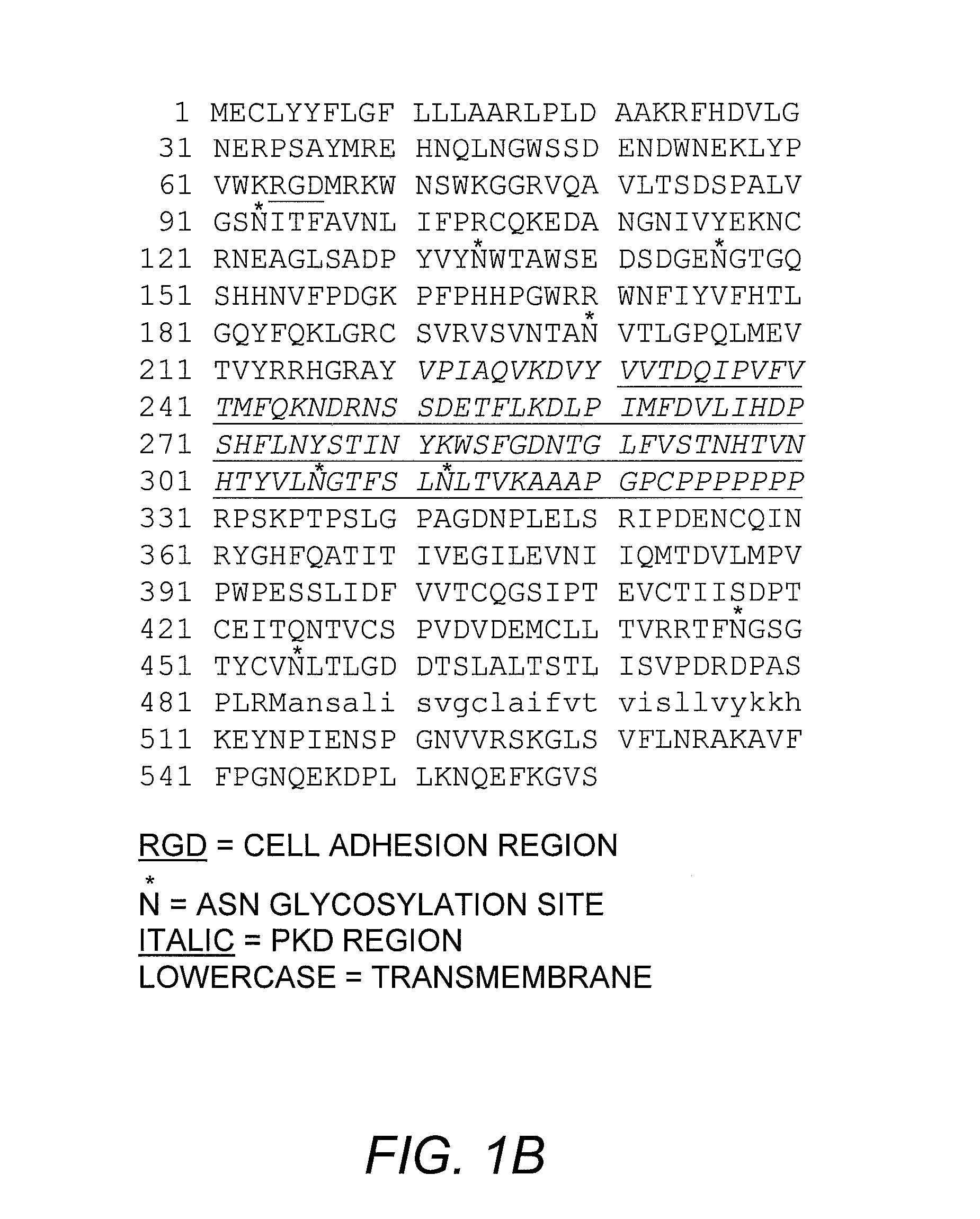
[0224] PCR was used to amplify the coding region of HGFIN, +60 / +1760 (GENBANK accession number AF322909). The following primer pairs were used in the PCR reaction: 5′-CGG GGT ACC ATG GAA TGT CTC TAC TA-3′ (SEQ ID NO:17) (upstream with KpnI linker) and 5′-CCG GAA TTC TCG AAA TTT AAG AAA CT-3′ (SEQ ID NO:18) (downstream with EcoRI linker). The HGFIN-specific sequences are underlined for both the upstream and downstream primers.

[0225] The amplified DNA fragment was cloned into pHAT10, hereafter referred as pHAT10-HGFIN. The vector was transformed into bacteria and HGFIN-HAT induced with IPTG. Induced bacterial cultures (20 ml) were sonicated in 2 ml of 100 mM Tris, pH 6.8, 4% SDS. After this, HGFIN was verified in the cell-free lysates by western blot analysis using 15 μg of total protein and rabbit anti-HAT. Details on the technique for western blot are described herein. The lysates that showed a band at the predicted size of...

example 3

ProteinChip Analyses for HGFIN-SP Interaction

[0226] Before studying the interaction between SP and HGFIN, each protein was profiled by the Surface Enhanced Laser Desorption / Ionization (SELDI) ProteinChip Array technology (Ciphergen Biosystems Inc., Fremont, Calif.). Normal phase (NP1) arrays were used for profiling and preactivated surface arrays (PSi) for HGFIN-SP interaction. For profiling studies, 2 μg of purified HGFIN or 2 μg of SP were spotted directly onto the NP1 arrays. Prior to adding of the proteins, chips were pre-wet with PBS. Arrays were incubated at room temperature until the protein was absorbed, which took approximately 5 to 10 minutes. After this, 0.5 μl of sinapinic acid (SPA) (Ciphergen Biosystems), diluted at 1:50 in 50% acetonitrile and 0.5% trifluoroacetic acid was added to the arrays. Chips were immediately analyzed using linear, time-lag focusing laser desorption / ionization SELDI-time-of-flight mass spectrometer (Model PBS II). Accurate mass was determined ...


PUM

PropertyMeasurementUnit
pHaaaaaaaaaa
temperatureaaaaaaaaaa
Tmaaaaaaaaaa

Abstract

The present invention discloses the cloning of a new cDNA, HGFIN, from stimulated bone marrow stromal cells that was retrieved with a probe specific for the neurokinin-1 (NK-1) receptor. The novel gene, HGFIN, encodes a protein receptor that is involved in the regulation of hematopoietic proliferation and differentiation. HGFIN is implicated in the treatment of hyperproliferative disorders, particularly bone and breast cancer, because it acts to suppress the proliferating cells.

Description

INTRODUCTION [0001] This application is a continuation-in-part application of U.S. patent application Ser. No. 10 / 463,106, filed Jun. 17, 2003, which is a continuation-in-part of U.S. patent application Ser. No. 10 / 039,272, filed Oct. 20, 2001, now U.S. Pat. No. 6,939,955, which claims priority to provisional patent application U.S. Ser. No. 60 / 241,881, filed Oct. 20, 2000, the disclosures of which are incorporated by reference in their entirety herein. [0002] This invention was made with government support by the following Public Health Service grants: HL-54973 and HL-57675 from the National Institute of Health and CA89868 from the National Cancer Institute. The government may own certain rights in the present invention.BACKGROUND OF THE INVENTION [0003] Bone Marrow is the major source of both lymphocytes (immune cells) and erythrocytes in the adult. Among the various cells that constitute the bone marrow are primitive hematopoietic pluripotent stem cells and progenitor cells. An i...

Claims


Application Information

Patent Timeline
Patent Type & Authority Applications(United States)
IPC IPC(8): A61K48/00A61P35/00C12N5/08A61K47/48C07K14/72
CPCC07K14/723A61K47/48269A61K47/642A61P35/00
Inventor RAMESHWAR, PRANELA
Owner RUTGERS THE STATE UNIV
Why Eureka
Try Eureka