Model-based registration of cardiac CTA and MR acquisitions

a technology of applied in the field of model-based registration of cardiac cta and mr acquisition, can solve the problems of limited spatial coverage and limited technique of each techniqu

Inactive Publication Date: 2003-05-13
SIEMENS MEDICAL SOLUTIONS USA INC
View PDF8 Cites 102 Cited by
  • Summary
  • Abstract
  • Description
  • Claims
  • Application Information

AI Technical Summary

Problems solved by technology

Although these techniques both provide useful information, each techniques may be limited by the by the type of information the techni

Method used


Image

  • Model-based registration of cardiac CTA and MR acquisitions
  • Model-based registration of cardiac CTA and MR acquisitions
  • Model-based registration of cardiac CTA and MR acquisitions

Examples


Embodiment Construction

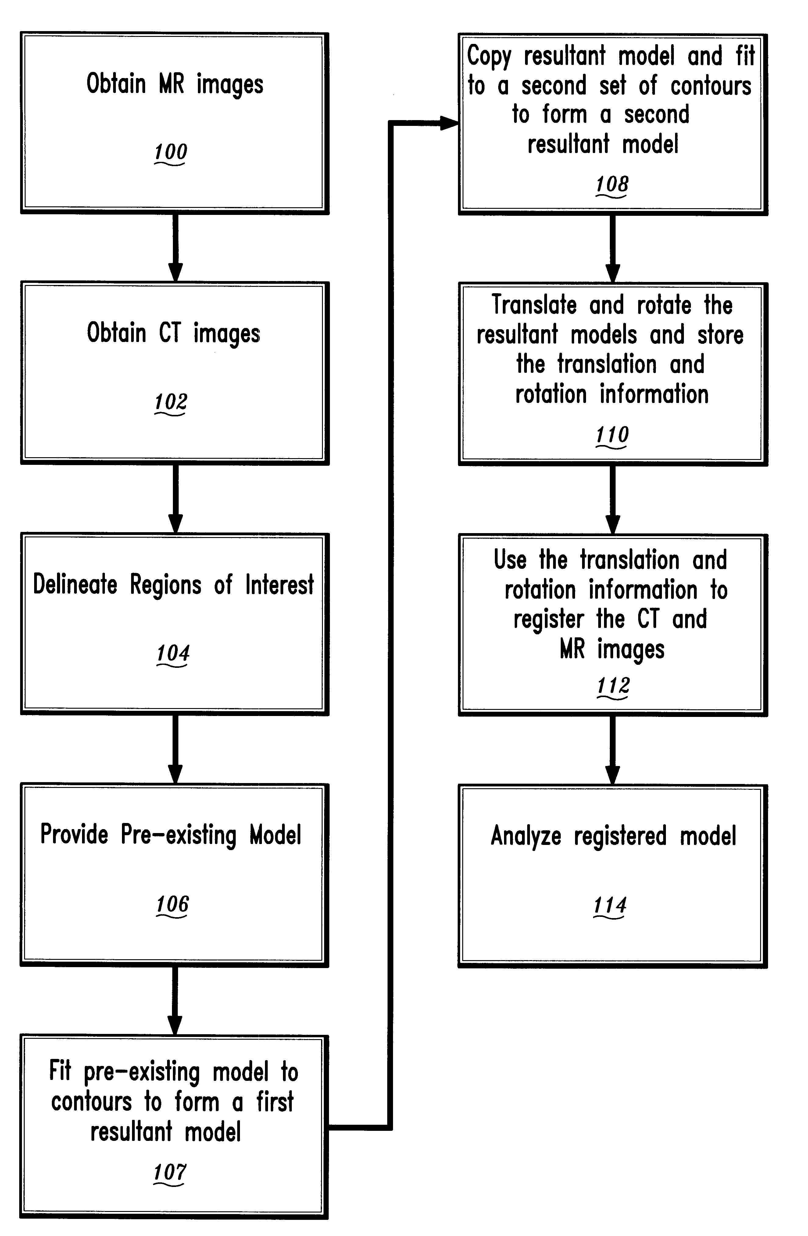
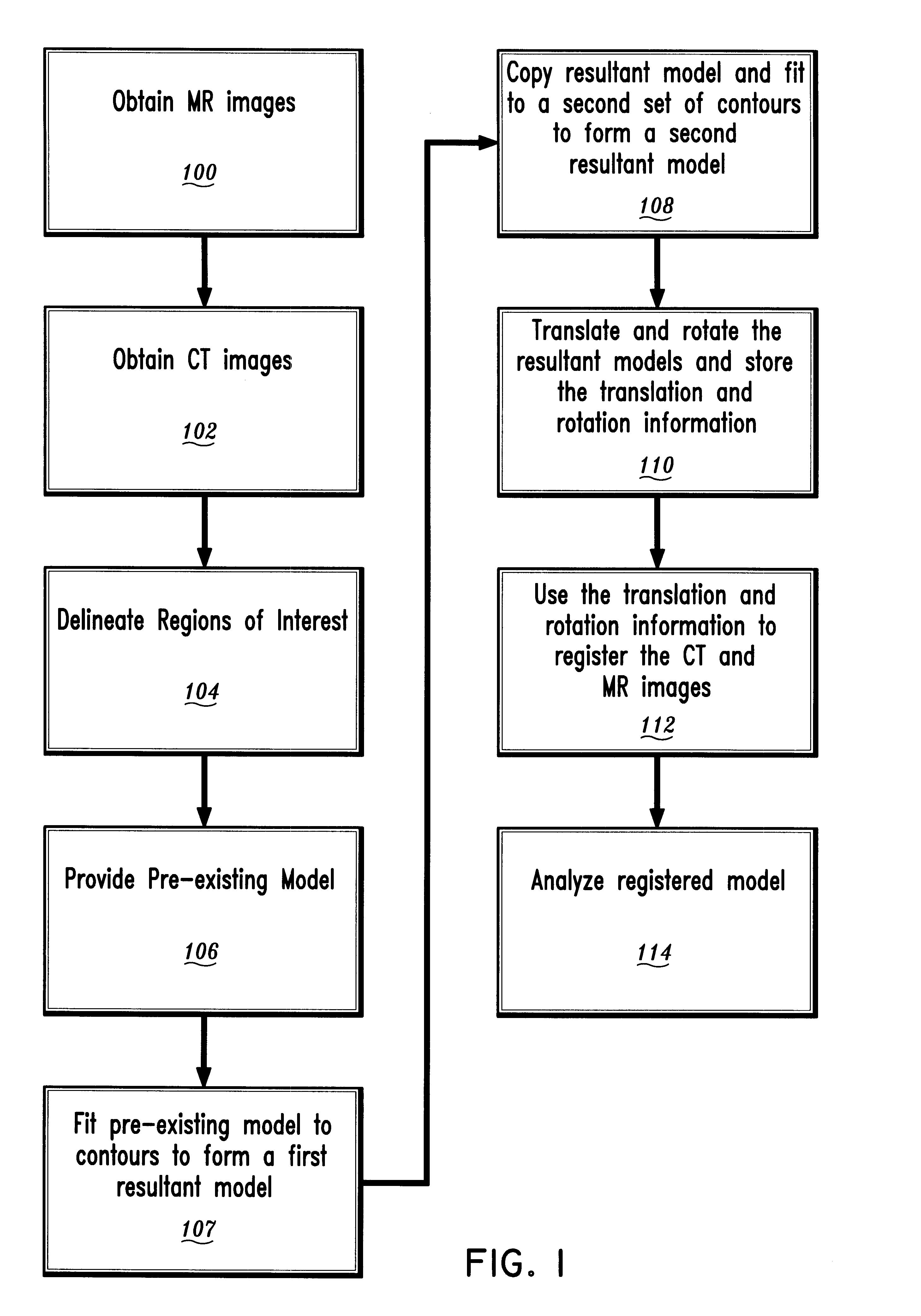
The present invention was employed to register CT and MR images of a human left ventricle. A patient with cardiovascular disease was imaged on a 1.5 T MR scanner (Symphony, Siemens Medical Systems, Erlangen, Germany) using a phased array body coil. A breath-hold 2D cine view-shared segmented FLASH sequence was used to evaluate wall motion. Short-axis 10 mm thick slices were obtained from the base to the apex of the heart with an in-plane interpolated image resolution equal to 1.17.times.1.17 mm.sup.2. Fifteen cardiac phases were acquired during the cardiac cycle at each position. Three long-axis views were also obtained with an interpolated image resolution equal to 1.25.times.1.25 mm.sup.2.

The patient was then imaged on a multi-slice CT scanner (SOMATOM Plus 4 Volume Zoom, Siemens Medical Systems, Erlangen, Germany). Retrospectively ECG-gated contrast-enhanced spiral scans were used to obtain CTA images of the coronary arteries. A 120 ml volume of Ultravist 240 (commercially availa...


PUM


Abstract

A method for registration of magnetic resonance (MR) and computed topography (CT) images, in accordance with the present invention includes providing MR images having a region of interest delineated by first contours and providing CT images having the region of interest delineated by second contours. A pre-existing model of the region of interest is also provided. The pre-existing model is fit to the first contours or the second contours to provide a first resultant model. The first resultant model is then copied to provide a copied model. The copied model is fit to the other of the first contours and the second contours to provide a second resultant model. By rotating and translating the first resultant model and the second resultant model, the first resultant model and the second resultant model are registered. The rotation and translation information are stored and applied to the images to provide registration between the MR images and the CT image

Description

1. Technical FieldThis disclosure relates to imaging and more particularly, to a method and apparatus for registering magnetic resonance and CT scan images.2. Description of the Related ArtMedical imaging of internal organs may be performed by non-invasive imaging procedures, such as computed topography (CT, or "Cat scan") and magnetic resonance (MR) imaging (MRI). These techniques provide useful information about internal organs which may be employed by medical personnel to make important health related decisions about a patients condition.Although these techniques both provide useful information, each techniques may be limited by the by the type of information the technique can provide. For example, CT imaging provides topographical images of internal structures of tissue. This means the shape or geometry of an internal organ can be determined using the CT procedure. MR imaging on the other hand provides functional information about the tissue comprising the organ or internal stru...

Claims


Application Information

Patent Timeline
IPC IPC(8): G06T3/00G06T7/00G01R33/32A61B5/055A61B6/03G06T1/00G06T19/00
CPCG06T3/4061G06T7/0038G06T7/0028G06T2207/30004G06T7/33G06T7/38
Inventor O'DONNELL, THOMASGUPTA, ALOKAHARON, SCHMUEL
Owner SIEMENS MEDICAL SOLUTIONS USA INC
Why Eureka
Try Eureka