System and method for providing information for detected pathological findings

a technology of pathological findings and information systems, applied in the field of business methods, can solve the problems of limited resources available to physicians to review, unnoticed and/or untreated pathology, etc., and achieve the effect of improving patient care quality

Inactive Publication Date: 2005-01-04
KAUFMAN SCOTT
View PDF6 Cites 31 Cited by
  • Summary
  • Abstract
  • Description
  • Claims
  • Application Information

AI Technical Summary

Benefits of technology

According to one embodiment, the invention is a method of providing information concerning pathological findings that allows a physician to be readily alerted to the existence of a pathology, which may include but is not limited to a disease, an injury and / or abnormality. The method involves receiving patient's demographic information, for example, name, sex, birth date, as well as a study description detailing the region of the patient's body to be imaged. During a magnetic resonance imaging procedure the patient is placed into an imaging volume of a magnet which generates a static magnetic field. Generally the procedure comprises of a predetermined scanning sequence based on the region of the body being scanned. Each scanning sequence is precisely timed, and directed to a series of pulse sequences for each data sampling window. Thus an imaging procedure for one region of the body may result in one or more scanned images.
Once the image is generated it is available for immediate viewing. Central controller can readily transmit the scanned image to a viewing console or monitor. The available image allows immediate interpretation by one skilled in the art of reading and interpreting radiographical images. Additionally, an operator may verify that the MRI scanner properly targeted the area being studied. If an interpreter is unavailable to read the image immediately, central controller may store the image along with the patient's demographic information in a large capacity storage device like a database to be retrieved at a later time.
Accordingly, the patient and the physician benefit from this method of providing pathological information evidenced from radiographical reports. The pathology is annotated for the doctor denoting exactly where the existing pathology is located. The annotated pictorial representation is viewable without the aid of a specialized viewer, and for doctors who are not well versed in interpreting an MRI image, the annotation readily identifies the type of pathology. The physician is now better equipped with the necessary information to provide a complete diagnosis for the patient. Both patient and the doctor will benefit from the increased quality patient care as pathologies are detected and confirmed sooner, thus either preventative or corrective treatment can be implemented as required.

Problems solved by technology

Unfortunately, unless a doctor is extremely vigilant and careful, a detected pathology may remain unnoticed and / or untreated until the patient's next scheduled visit.
This is because the physician may have limited resources available to review incoming reports in order to satisfy the demands of their medical practice at the time.

Method used


Image

  • System and method for providing information for detected pathological findings
  • System and method for providing information for detected pathological findings
  • System and method for providing information for detected pathological findings

Examples


Embodiment Construction

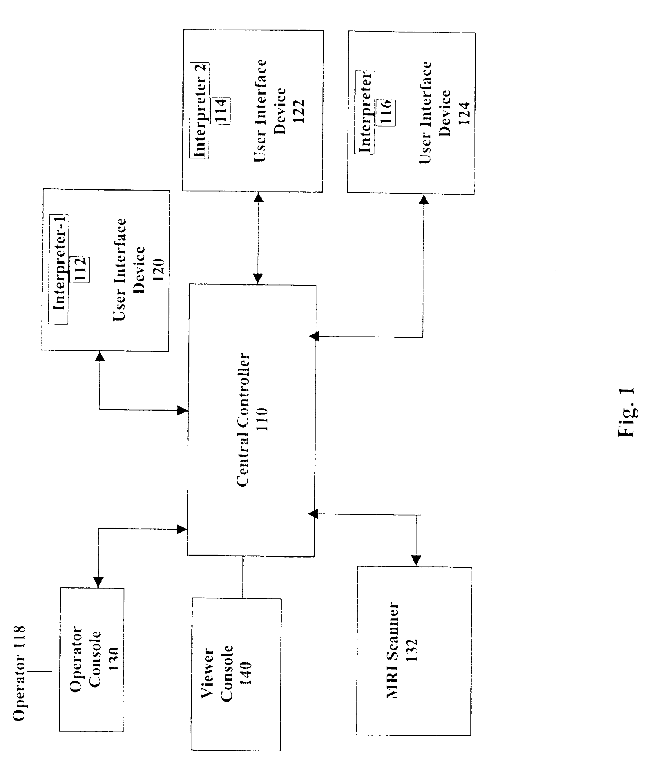
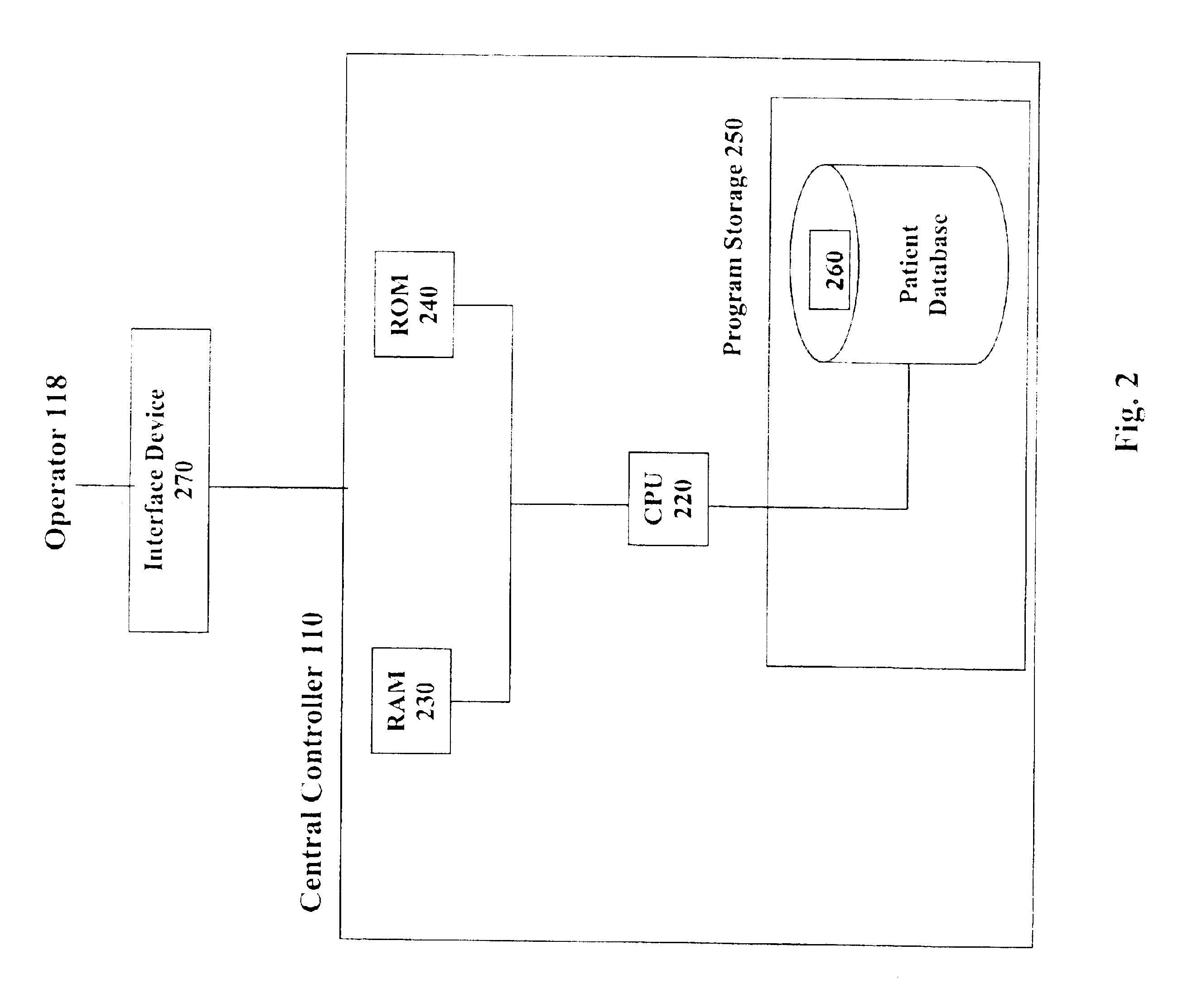
FIG. 1 illustrates an example of the functional components of a system in accordance with the present invention. As described below, the system and method allows for the provision of information concerning pathological findings evidenced by radiographical reports. The system incorporates a number of functional components. In the illustrated embodiment, the system comprises of a central controller 110 electronically coupled to a MRI scanner 132, operator console 130, and a viewer console 140. The system may also have interpreters 112, 114, 116 linked in a network wherein the interpreters 112, 114, 116 can readily access the patient's images at their convenience for reading and interpretation using interface devices 120, 122, 124.

As is well known, the MRI scanner 132 comprises, in general, of a current supply device and a measuring station, which includes magnets for the creation of high frequency magnetic pulses. The magnet of the MRI scanner 132 is preferably in an open configuratio...


PUM


Abstract

A system and method of providing information concerning pathological findings evidenced by radiographical reports. A medical care provider may request a radiographical image, in particular, an MRI image of a region of the body. The region of the patient's body is scanned and the images are read and interpreted to determine if there is a positive finding of a pathology. If a pathology is detected, the generated scanned images are reviewed for a radiographical image that best depicts the detected pathology and the image is selected for further annotation. The image is then annotated to highlight and more fully describe and delineate the pathology thus alerting the medical care provider of the pathology's existence. In addition, the report may be further delineated with an identifying mark informing the referring physician of a positive finding of a pathology.

Description

FIELD OF THE INVENTIONThis invention generally relates to a business method and more particularly, to a system and method of providing information concerning pathological findings evidenced by radiographical reports.BACKGROUND OF INVENTIONMagnetic Resonance Imaging (MRI) is a well-known diagnostic tool that is primarily used by doctors to view internal images of the human body from various angles without the use of x-rays or invasive procedures. MRIs provide the added benefit of producing imaging results in a relatively short period of time. A MRI image also provides basic biomedical and anatomical information in such precise detail that it facilitates early diagnosis of injuries, diseases or abnormalities. When a patient is scanned, internal images of the body are produced by nuclear magnetic resonance. The images are represented in contrast by light and dark areas depending on the density and structure of the region of the body being scanned. The scanned image is then interpreted ...

Claims


Application Information

Patent Timeline
Patent Type & Authority Patents(United States)
IPC IPC(8): A61B6/00G06F19/00A61B5/055G16H30/40
CPCA61B6/00A61B6/461G06F19/345G06F19/327G06F19/3406A61B6/468A61B5/055G06F19/321G16H40/63G16H40/20G16H50/20G16H30/40
Inventor KAUFMAN, SCOTT
Owner KAUFMAN SCOTT
Why Eureka
Try Eureka