Compound image display system and method

a compound image and display system technology, applied in tomography, instruments, applications, etc., can solve problems such as undesirable noise in the storage image, and achieve the effect of reducing noise and increasing speckle reduction

Inactive Publication Date: 2005-03-29
SIEMENS MEDICAL SOLUTIONS USA INC
View PDF8 Cites 18 Cited by
  • Summary
  • Abstract
  • Description
  • Claims
  • Application Information

AI Technical Summary

Benefits of technology

The compounded sub-set image is associated with reduced noise as compared to the image absent compounding and more closely resembles an area of the extended field of view image. Where a plurality of images are associated with a rotated transducer and scan planes or where a sector or Vector® scan pattern is used, the compounded sub-set provides spatial compounding associated with different angles of incidence to the region of interest, increasing speckle reduction.

Problems solved by technology

The stored image may include undesirable noise and / or look different than the corresponding region of the extended field of view image.

Method used


Image

  • Compound image display system and method
  • Compound image display system and method
  • Compound image display system and method

Examples


Embodiment Construction

To provide diagnostic information, information for an extended field of view is obtained. The information includes two or more frames of data associated with scans of a patient corresponding to two or more transducer positions, respectively. The frames of data are compounded together to reduce speckle. Various images are provided using this information, such as an extended field of view image corresponding to the compounded frames of data, a compounded image corresponding to a sub-set of the extended field of view compounded information or an image corresponding to one of the frames of data absent the compounded for forming the extended field of view. These images and comparison between these images assists in medical diagnosis.

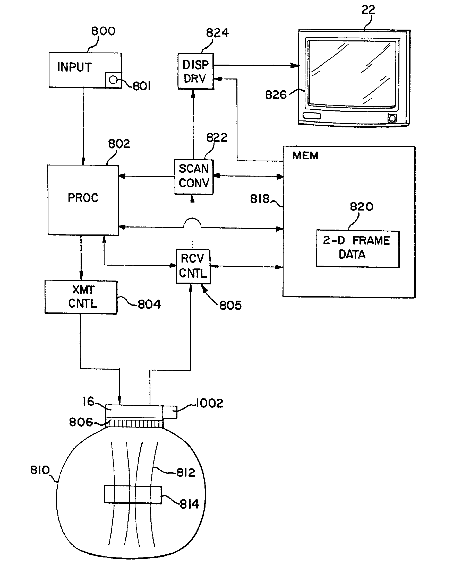
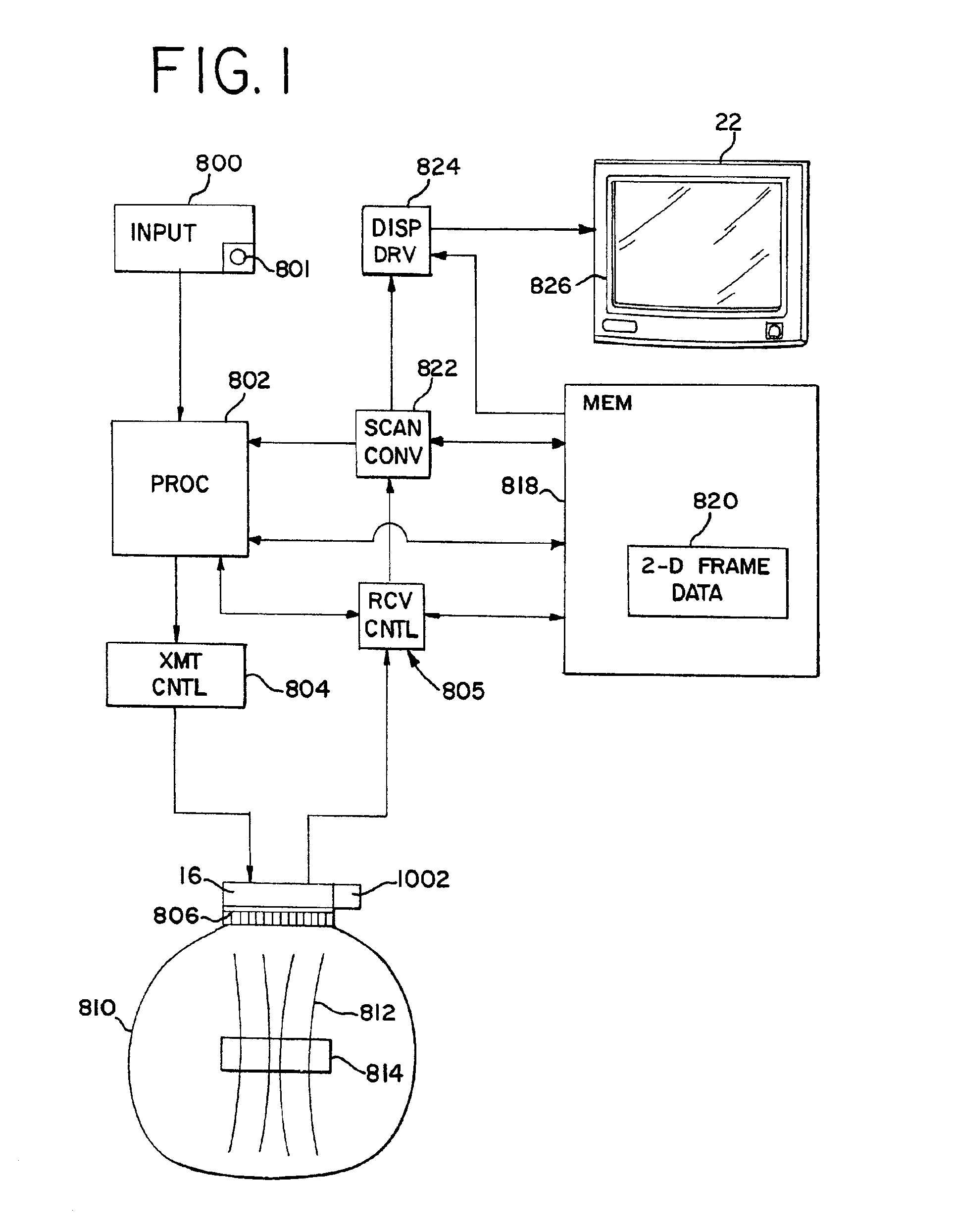
FIG. 1 illustrates an ultrasonic imaging system according to one embodiment. The system includes an input 800, a processing system 802, a transmission control 804, a transducer 16 with transducer elements 806, a display 22, a receive control 805, a scan conve...


PUM


Abstract

A method and system for displaying an image associated with an extended field of view is provided. One image comprises a compounded sub-set of the extended field of view. Other images may include an extended field of view image or an image absent compounding from the extended field of view image. The compounded sub-set image is associated with reduced noise as compared to the image absent compounding and more closely resembles an area of the extended field of view image. Where plurality of images are associated with a rotated transducer or scan planes or where a sector or Vector® scan pattern is used, the compounded sub-set provides spatial compounding associated with different angles of incidence to the region of interest, increasing speckle reduction.

Description

BACKGROUNDThis invention relates to spatial compound imaging. In particular, displays associated with images compounded from scans of different areas are provided.Extended field of view (i.e., panoramic) imaging provides compound imaging. U.S. Pat. Nos. 5,566,674, 5,575,286, 5,782,766 and 6,014,473 describe various techniques for extended field of view imaging. In general, a transducer is moved along an azimuthal dimension to acquire frames of data associated with different transducer positions within an imaging plane. One of various techniques are used to determine the position of each frame of data relative to other frames of data. The frames of data are spatially aligned and combined to form the extended field of view image. The combination may include compounding data representing same locations.Typically, the extended field of view image is shown on a display. Associated images may also be generated. In one configuration shown in U.S. Pat. No. 5,782,766, a region of the extende...

Claims


Application Information

Patent Timeline
Patent Type & Authority Patents(United States)
IPC IPC(8): A61B8/00G09G5/00A61B8/14G01S7/52G01S15/89
CPCA61B8/14G01S15/8995G01S7/52074A61B8/5238
Inventor TIRUMALAI, ARUN P.SUTCLIFFE, PATRICK L.
Owner SIEMENS MEDICAL SOLUTIONS USA INC
Why Eureka
Try Eureka