Suture anchor

a technology of suture anchors and anchors, which is applied in the field of suture anchors, can solve the problems of joint function loss to a greater or lesser degree, soft tissue damage,

Active Publication Date: 2005-08-23
DEPUY SYNTHES PROD INC
View PDF16 Cites 312 Cited by
  • Summary
  • Abstract
  • Description
  • Claims
  • Application Information

AI Technical Summary

Problems solved by technology

Frequently, as a result of trauma caused by an accident or overloading of a joint during sports activities, the soft tissue may become damaged.
Such injuries tend to result in loss of joint function to a greater or lesser degree, and typically require some sort of surgical intervention and remediation to provide normal joint function.

Method used


Image

  • Suture anchor
  • Suture anchor
  • Suture anchor

Examples


example 1

[0058]A patient is prepared for arthroscopic surgery for a repair of a soft tissue injury in the patient's rotator cuff in the following manner. The patient is placed in a lateral position, or in a semi-sitting (beach chair) position, on a standard operating room table. After skin preparation and draping, full exposure of the coracoid process anteriorly and the entire scapula posteriorly is obtained. A standard posterior gleno-humeral arthroscopy portal is employed. An 18 gauge spinal needle is inserted toward the coracoid process anteriorly, in order to enter the gleno-humeral joint. The joint is inflated with sterile saline, and free outflow of fluid is confirmed as the inflating syringe is removed. Prior to making all portal incisions, the skin and subcutaneous tissue are infiltrated with 1% Lidocaine with Epinephrine, in order to reduce bleeding from the portals. The arthroscope cannula is inserted in the same direction as the spinal needle into the joint and free outflow of flu...

example 2

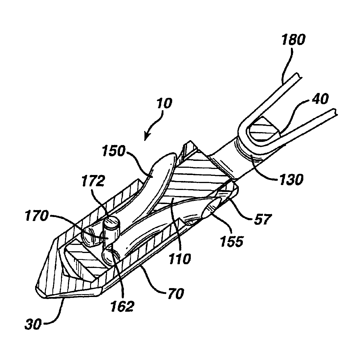
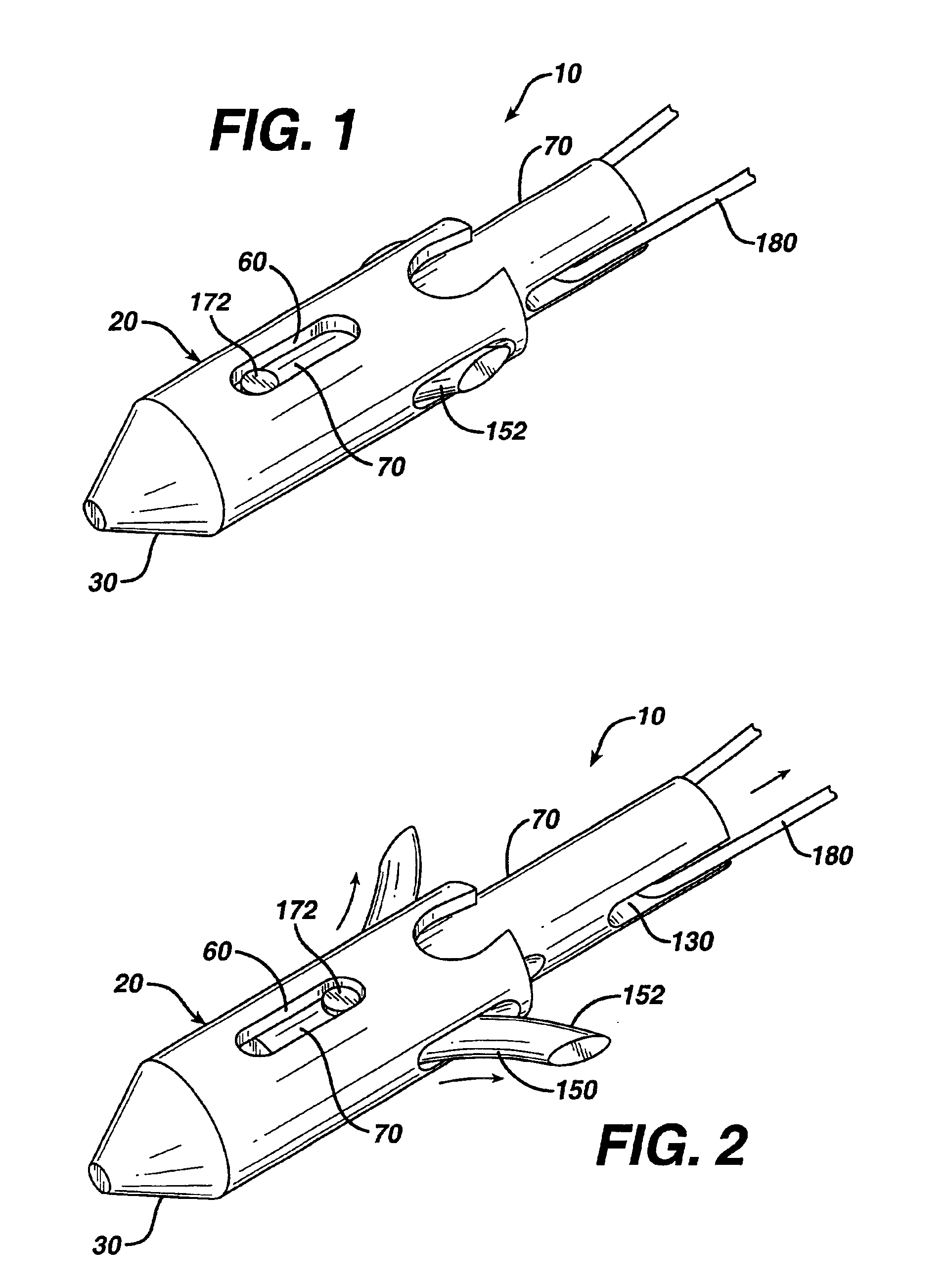
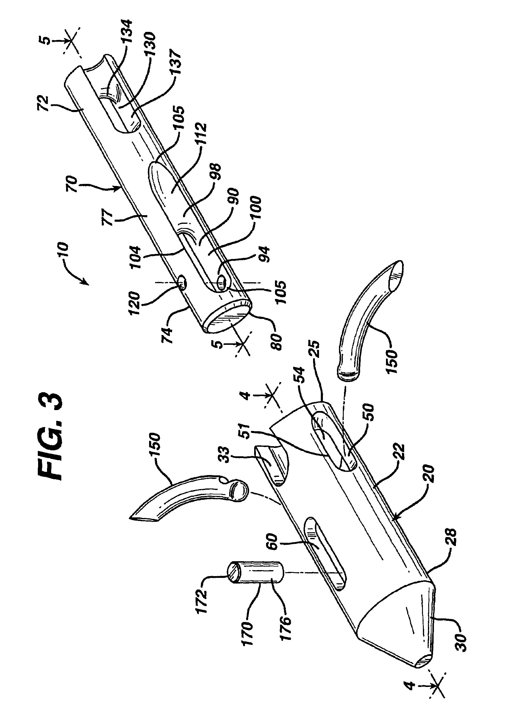
[0059]A second patient undergoes a similar surgical procedure to that described in Example 1. During the procedure, and after emplacement and engagement of suture anchor 10 in the drilled blind bore hole, it is necessary to remove the deployed anchor 10. The surgeon removes anchor 10 in the following manner. The insertion device 200 is positioned adjacent to the proximal end of the suture anchor 10, in alignment with axis of the device. The insertion device 200 is reattached to the anchor by pressing the distal tip of the inserter 200 onto the proximal end of the suture anchor 10 in openings 31. Continued insertion pressure is applied so that as the suture anchor 10 moves deeper into the drilled hole, the engagement members 150 retract into the slots 50 of outer member 20 and into cavity 90 of actuation member 70 of the suture anchor 10. The suture anchor 10 is then removed from the drill hole by removing the inserter / anchor assembly through the cannula.

[0060]An alternate embodiment...


PUM


Abstract

A suture anchor for affixing soft tissue to bone. The anchor has a hollow outer member, and an actuation member slidably mounted within the outer member. The actuation member has at least two engagement members that are pivotally mounted to the actuation member. Proximal movement of the actuation member with respect to the outer member causes the engagement members to move outward to a deployed position, wherein the engagement members are deployed in bone. A surgical suture is mounted to the anchor.

Description

TECHNICAL FIELD[0001]The field of art to which this invention pertains is medical devices and surgical procedures for repairing soft tissue injuries, more specifically, suture anchors and methods of using these suture anchors to affix soft tissue to bone.BACKGROUND OF THE INVENTION[0002]Suture anchor devices are known in the art of arthroscopy and orthopedic surgery for use in repairing soft tissue injuries in joints. A joint, such as the knee, the elbow, or the shoulder, contains tissue such as ligaments, tendons and cartilage, which allows the joint to function. For example, the soft tissue permits joint articulation, and the transmission and absorption of forces. Frequently, as a result of trauma caused by an accident or overloading of a joint during sports activities, the soft tissue may become damaged. It is not unusual in a sports related injury to have tearing of cartilage or the separation of a tendon or ligament from a bone surface. Such injuries tend to result in loss of j...

Claims


Application Information

Patent Timeline
Patent Type & Authority Patents(United States)
IPC IPC(8): A61B17/04A61B17/72A61B17/68A61B17/56
CPCA61B17/0401A61F2/0811A61B17/7266A61B2017/0409A61B2017/0412A61B2017/0414A61B2017/0435A61F2002/0888
Inventor LIZARDI, JOSE E.HOWE, JONATHAN EMERSONMCALISTER, GARYJENSEN, KENNETH L.PERKINS, DANIEL A.
Owner DEPUY SYNTHES PROD INC
Why Eureka
Try Eureka