Implantable medical electrical stimulation lead fixation method and apparatus

a technology of medical electrical stimulation and lead fixation, which is applied in the field of method and apparatus, can solve the problems of low success rate of surgical procedures, inability to reverse, and insufficient medical treatment of disease states,

Inactive Publication Date: 2006-02-14
MEDTRONIC INC +1
View PDF87 Cites 344 Cited by
  • Summary
  • Abstract
  • Description
  • Claims
  • Application Information

AI Technical Summary

Benefits of technology

[0025]The present invention recognizes and provides a solution to the problems associated with implanting and maintaining electrical leads in body tissue, particularly muscle tissue to maintain one or more lead electrode in relation to a particular body site, through use of minimally invasive implantation techniques. In a preferred embodiment, the present invention maintains one or more sacral nerve stimulation electrode in close proximity or casual contact with discrete nerve fibers of the sacral nerves by providing a unique solution that allows minimally invasive, percutaneous, implantation near to, but avoiding compressive contact with, the sacral nerves and fixation of the lead body in subcutaneous tissues. Additionally, the invention provides apparatus and a method of implanting a medical electrical stimulation lead through the subcutaneous tissue and foramen for stimulating a bundle of nerve fibers of the sacral nerve anterior to the sacral nerve opening through the sacrum.
[0029]In another embodiment, each of the M tine elements comprises a tine mounting band encircling the lead body with the N tines extending from respective attached tine ends disposed apart from one another, preferably at equal spacing, around the tine mounting band. The N tines have a tine thickness that enables folding of the tines against the lead body in the space between the tine mounting band and the adjoining proximal tine mounting band.
[0034]The tines engage subcutaneous muscle layers that are penetrated by the introducer, not a pre-existing anatomical structure or a subcutaneously tunneled space overlying muscle layers. The tines are designed specifically to engage the muscle tissue and are relatively short and stiffer than the soft, pliant tines or projections on endocardial leads that get entangled in the trabeculae. Short, stiff tines provide better acute fixation in the muscle layers, and relatively harp, non-tapered, tine edges are provided to “dig into” the muscle.
[0035]In an alternative embodiment, one or more or all of the P stimulation electrodes is preferably formed of a flexible elongated mesh or wire coil electrodes that can bend somewhat to fit through a foramen to locate the elongated electrode optimally with respect to a sacral nerve. The elongated electrode surface area of the mesh or coiled wire electrode creates a wider electric field which allows the lead to be placed in a less precise or gross manner while still providing adequate electrical stimulation to the sacral nerve.
[0036]Accordingly, the present invention advantageously provides a unique implantable medical neurostimulation lead that provides adequate stimulation of the sacral nerves for control of incontinence and other pelvic floor disorders with the sacral nerves and with less sensitivity to placement and dislodgement. The unique lead simplifies the implant procedure and reduces or eliminates the need to reprogram the implantable pulse generator stimulation levels or re-open the patient to re-position the stimulation electrodes since the electrodes are less likely to move.
[0037]The implantation method for implanting the lead of the present invention allows more rapid placement of the electrodes for the treatment of incontinence whereby the lead is placed near the sacral nerves. Implanting the medical electrical lead near the sacral nerves with less specificity as to location near the sacral nerves reduces the time for implantation. Currently, the implantation procedure for existing medical electrical leads stimulating the sacral nerve fibers takes approximately 20–60 minutes. The present invention allows for implantation near the sacral nerve bundle and reduces the time for implantation to approximately 5–10 minutes.

Problems solved by technology

Many of the pharmaceuticals do not adequately resolve the issue and can cause unwanted side effects, and a number of the surgical procedures have a low success rate and are not reversible.
These solutions have drawbacks well known to those skilled in the art.
In addition, some disease states do not have adequate medical treatments.
A problem associated with implantation of permanent and temporary neurostimulation leads involves maintaining the discrete ring-shaped electrode(s) in casual contact, that is in location where slight contact of the electrode with the sacral nerve may occur or in close proximity to the sacral nerve to provide adequate stimulation of the sacral nerve, while allowing for some axial movement of the lead body.
Typically, physicians spend a great deal of time with the patient under a general anesthetic placing the leads due to the necessity of making an incision exposing the foramen and due to the difficulty in optimally positioning the small size stimulation electrodes relative to the sacral nerve.
The patient is thereby exposed to the additional dangers associated with extended periods of time under a general anesthetic.
As can be appreciated, unintended movement of any object positioned proximate a nerve may cause unintended nerve damage.
But, too close or tight a contact of the electrode with the sacral nerve can also cause inflammation or injury to the nerve diminishing efficacy and possibly causing patient discomfort.
These surface treatments or geometries provide some acute fixation against the subcutaneous tissues, but they are necessarily insufficient to resist intentional retraction of the lead to remove it upon cessation of temporary stimulation.
However, it was found in practice that the canted tip is unnecessary and that only three, much shorter, tines in the 270° arc or four tines spaced apart by 90° in a common circumference like a ventricular tined lead, are sufficient.
The rows of tines shown in the '843 patent are necessarily closely spaced because of the small area of trabeculae in the right atrium, and more proximal tines simply typically do not engage anything and make it difficult to lodge any of the tines in the interstitial spaces.
Furthermore, it is less critical to maintaining exact positioning of the sense electrodes of such a subcutaneous cardiac monitor than maintaining stimulation electrodes in the implanted positions for cardiac or nerve or muscle stimulation because minute stimulation electrode movement can decrease efficacy and increase battery energy consumption or fail.

Method used


Image

  • Implantable medical electrical stimulation lead fixation method and apparatus
  • Implantable medical electrical stimulation lead fixation method and apparatus
  • Implantable medical electrical stimulation lead fixation method and apparatus

Examples


Embodiment Construction

and the preferred embodiments follow, after a brief description of the drawings, wherein additional advantages and features of the invention are disclosed.

[0039]This summary of the invention has been presented here simply to point out some of the ways that the invention overcomes difficulties presented in the prior art and to distinguish the invention from the prior art and is not intended to operate in any manner as a limitation on the interpretation of claims that are presented initially in the patent application and that are ultimately granted.

BRIEF DESCRIPTION OF THE DRAWINGS

[0040]Preferred embodiments of the invention are illustrated in the drawings, wherein like reference numerals refer to like elements in the various views, and wherein:

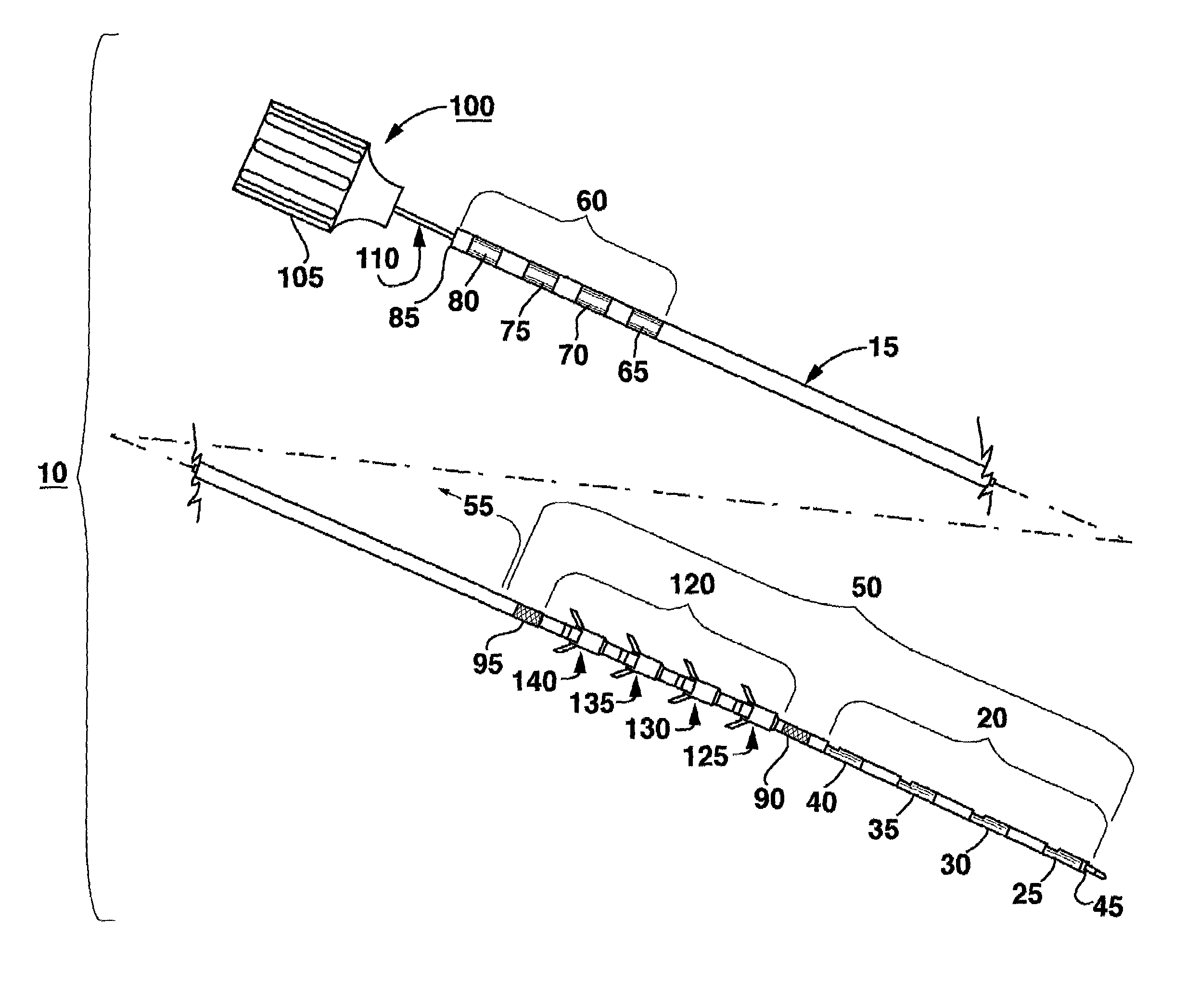
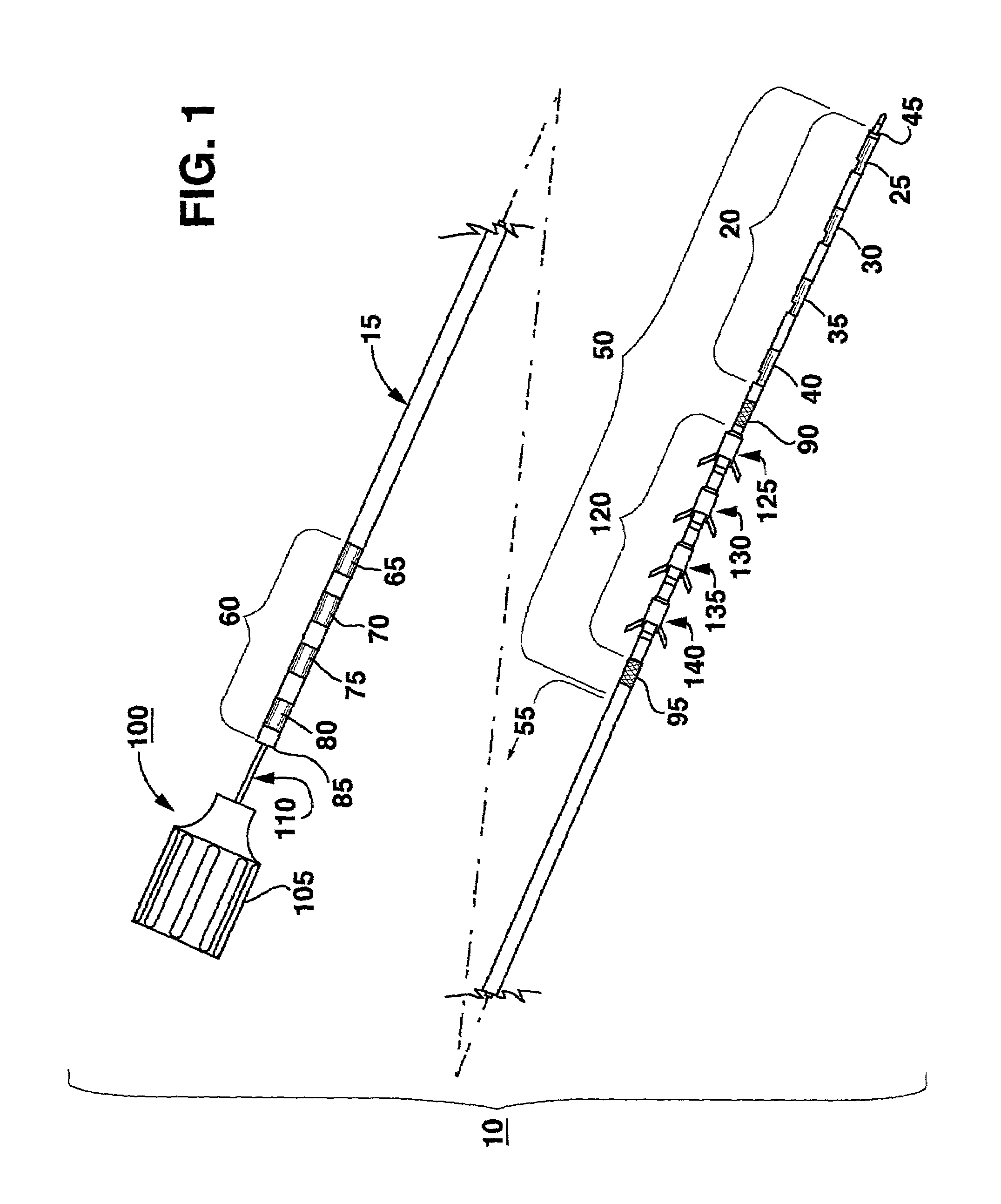
[0041]FIG. 1 is a plan view of one embodiment of sacral nerve stimulation lead of the present invention having a tine element array and stimulation electrode array in a distal portion of the lead body;

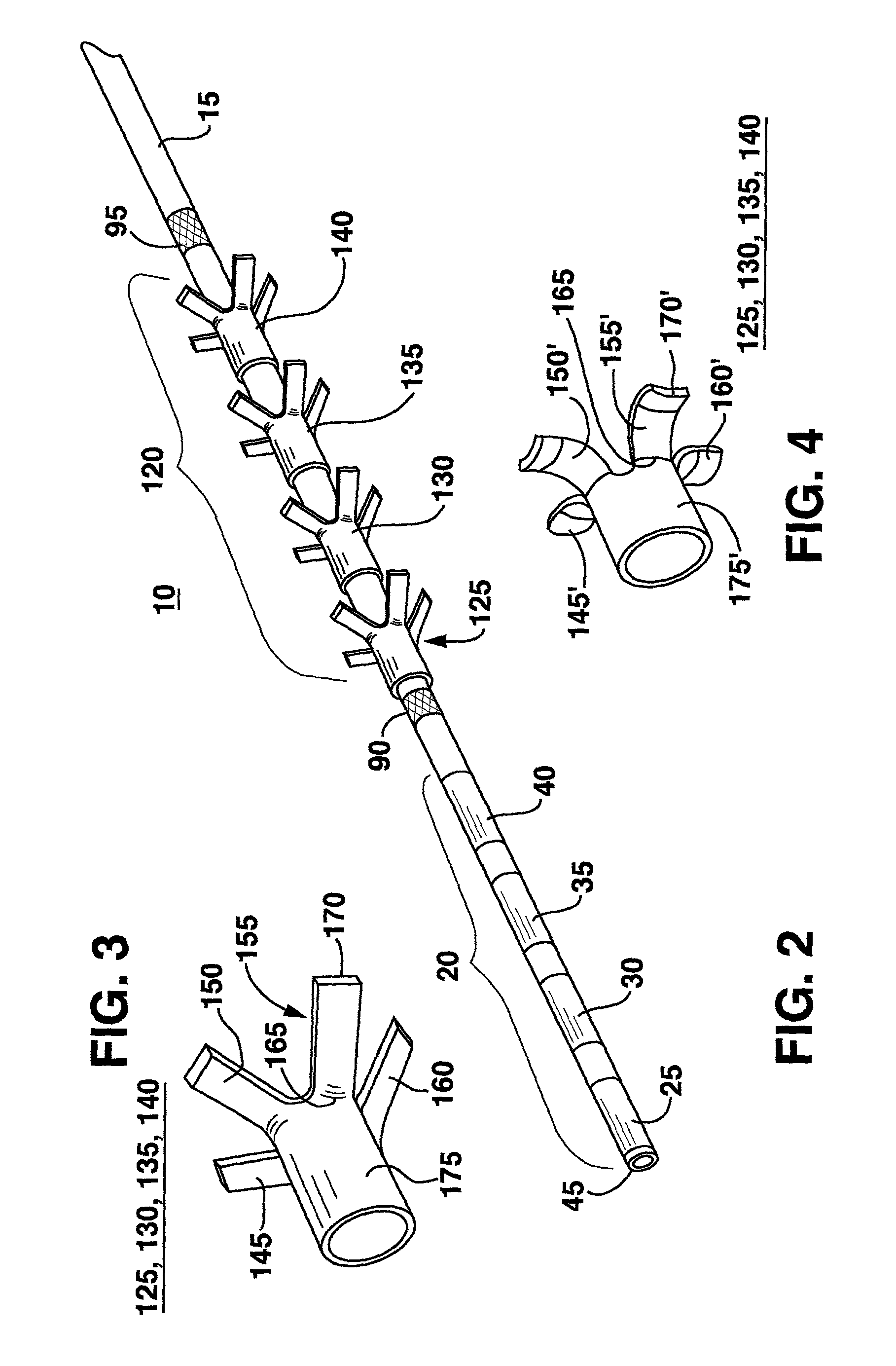
[0042]FIG. 2 is an expanded perspective vie...


PUM


Abstract

An implantable medical electrical lead particularly for stimulation of the sacral nerves comprises a lead body extending between a distal end and a proximal end, and the distal end having at least one electrode of an electrode array extending longitudinally from the distal end toward the proximal end. The lead body at its proximal end may be coupled to a pulse generator, additional intermediate wiring, or other stimulation device. A fixation mechanism is formed on or integrally with the lead body proximal to the electrode array that is adapted to be implanted in and engage subcutaneous tissue, particularly muscle tissue, to inhibit axial movement of the lead body and dislodgement of the stimulation electrodes. The fixation mechanism comprises a M tine elements arrayed in a tine element array along a segment of the lead proximal to the stimulation electrode array. Each tine element comprises at least N flexible, pliant, tines, each tine having a tine width and thickness and extending through a tine length from an attached tine end to a free tine end. The attached tine end is attached to the lead body from a tine attachment site and supports the tine extending outwardly of the lead body and proximally toward the lead proximal end. The M×N tines are adapted to be folded inward against the lead body when fitted into and constrained by the lumen of an introducer such that the tine free ends of more distal tines of more distal tine elements are urged toward or alongside the attached tine ends of the adjacent more proximal tines of more proximal tine elements, and the folded tines do not overlap one another.

Description

[0001]This application claims the benefit of Ser. No. 60 / 316,582 filed on Aug. 31, 2001.BACKGROUND OF THE INVENTION[0002]1. Field of the Invention[0003]This invention relates generally to a method and apparatus that allows for stimulation of body tissue, particularly sacral nerves. More specifically, this invention relates to an implantable medical electrical lead having at least one stimulation electrode adapted to be implanted near the sacral nerves for stimulation of a bundle of sacral nerve fibers and a fixation mechanism for providing chronic stability of the stimulation electrode and lead. Moreover, this invention relates to the method of implantation and anchoring of the medical electrical lead electrodes in operative relation to a selected sacral nerve to allow for stimulation.[0004]2. Description of Related Art[0005]Pelvic floor disorders such as, urinary incontinence, urinary urge / frequency, urinary retention, pelvic pain, bowel dysfunction (constipation, diarrhea), erecti...

Claims


Application Information

Patent Timeline
Patent Type & Authority Patents(United States)
IPC IPC(8): A61N1/05
CPCA61N1/0558A61N1/0551
Inventor SWOYER, JOHN MATTHEWCARLTON, KEITHGERBER, MARTIN T.MAMO, GEORGE J.SPINELLI, MICHELEHARTLE, STEVEN DAVID
Owner MEDTRONIC INC
Why Eureka
Try Eureka