Method and system for processing vascular radiographic images which have been reconstructed by three-dimensional modelling

a three-dimensional modelling and radiographic image technology, applied in image enhancement, image analysis, instruments, etc., can solve the problem of relatively difficult interpretation of the image obtained

Inactive Publication Date: 2006-05-16
GE MEDICAL SYST GLOBAL TECH CO LLC
View PDF17 Cites 8 Cited by
  • Summary
  • Abstract
  • Description
  • Claims
  • Application Information

AI Technical Summary

Problems solved by technology

a reconstructed model known as an “opacified” model, identifying the vascular elements, the calcified elements and the prosthe...

Method used


Image

  • Method and system for processing vascular radiographic images which have been reconstructed by three-dimensional modelling
  • Method and system for processing vascular radiographic images which have been reconstructed by three-dimensional modelling
  • Method and system for processing vascular radiographic images which have been reconstructed by three-dimensional modelling

Examples


Embodiment Construction

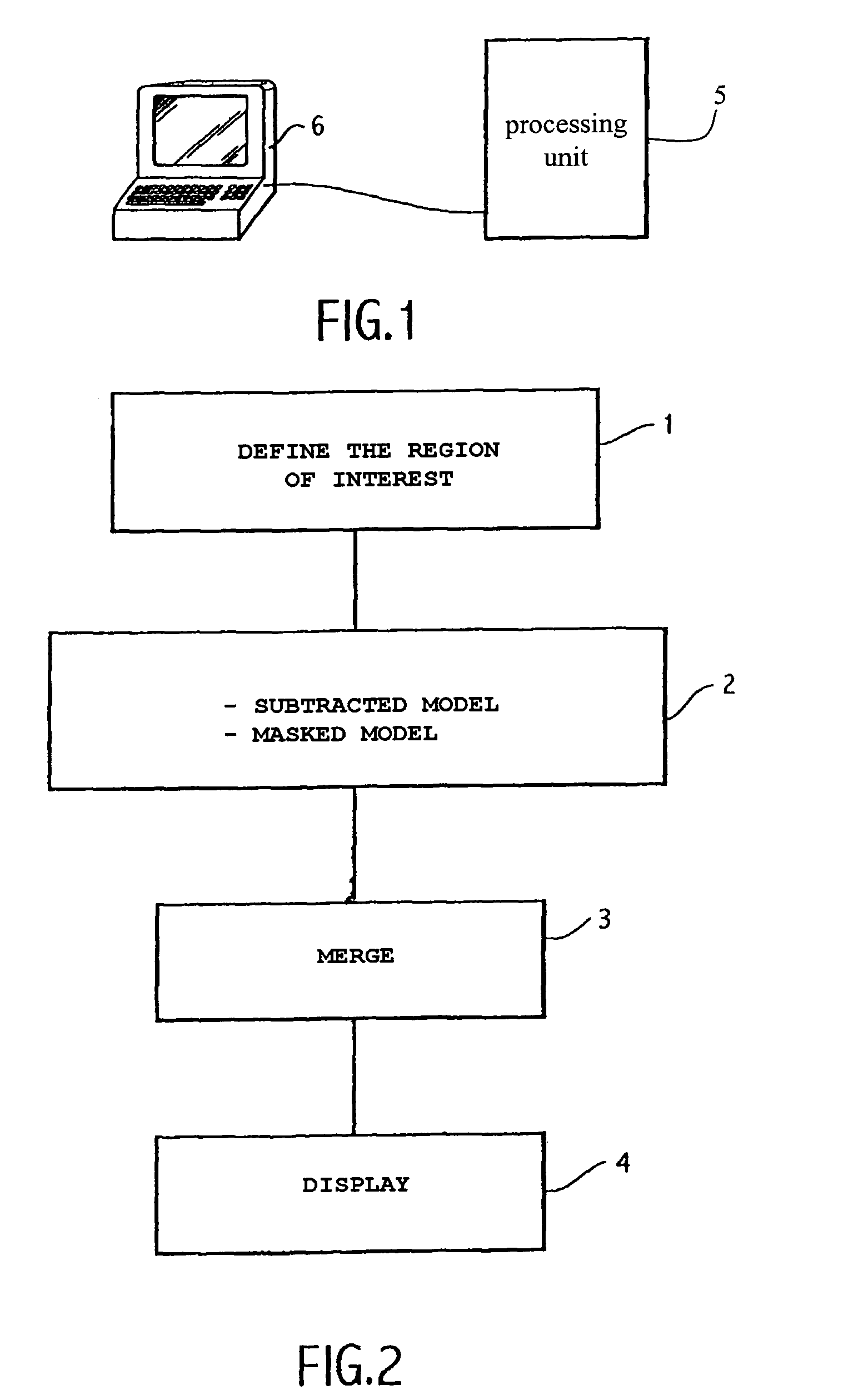
[0019]In this embodiment, it is assumed that there is available a set of two-dimensional angiography images obtained around a given anatomical region of a patient and from these images it is possible to reconstruct three-dimensional models of the anatomical region and, in particular, subtracted and masked three-dimensional models. These angiography images may, for example, be obtained by x-ray fluoroscopy, etc. The images are stored and processed in a processing unit 5, which is connected to interface means 6 which, in particular, allow the radiography images to be displayed (FIG. 1).

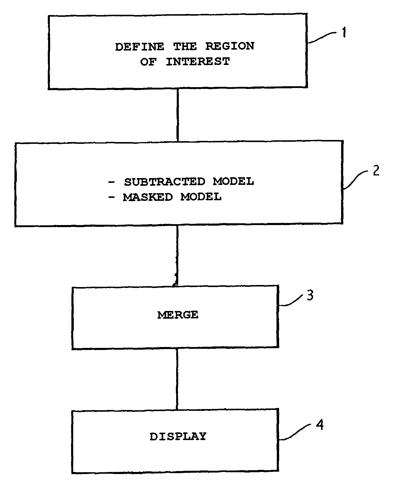
[0020]In an embodiment of the processing may be carried out in four successive steps.

[0021]In a first step (step 1 in FIG. 2), the user defines a volume which constitutes the region of interest. The region of interest will, in particular, preferably be defined so that it contains only a limited number of bones while at the same time fully including the portion or portions of blood vessel the user wishes...


PUM


Abstract

Method and system for processing vascular radiography images which have been reconstructed by three-dimensional modelling, in which: from this three-dimensional modelling there is determined a three-dimensional model known as the masked model which features the calcified elements and the prosthetic elements, but not the vascular elements; a three-dimensional model known as the subtracted model, which features the vascular elements alone, is determined; these two models are merged, weighting their voxels so as to increase the contrast between the images of the masked model and the images of the subtracted model; and summing the voxels thus weighted.

Description

CROSS-REFERENCE TO RELATED APPLICATIONS[0001]The entire contents of French Patent Application No. 0100737 filed Jan. 19, 2001 are hereby incorporated by reference.BACKGROUND OF THE INVENTION[0002]The present invention is directed to a method and system for processing radiographic images which have been reconstructed by three-dimensional modelling. In particular, the present invention is applicable to vascular images which includes an implanted element, such as a prosthesis.[0003]There are known numerous three-dimensional imaging methods and systems which take two-dimensional images obtained, for example, by X-ray fluoroscopy, to produce 3D models of an object, i.e., a patient that it is desired to be observe. In particular, there are known 3D angiography systems which, by X-ray fluoroscopy, reconstruct 3D models of vessels on which a procedure is to be carried out, for example in order to treat arterial stenosis. In 3D angiography, there are three complementary types of image likely...

Claims


Application Information

Patent Timeline
IPC IPC(8): G06K9/00G06T5/00G06T5/50
CPCG06T5/50G06T5/002G06T5/008G06T2207/30101G06T2207/10121G06T2207/20221G06T2200/04
Inventor LAUNAY, LAURENTBETTING, FABIENNEKNOPLIOCH, JEROME
Owner GE MEDICAL SYST GLOBAL TECH CO LLC
Why Eureka
Try Eureka