Assay system for simultaneous detection and measurement of multiple modified cellular proteins

a technology of cellular protein and detection system, which is applied in the field of simultaneous detection and measurement of multiple modified cellular proteins, can solve the problems of limited detection and measurement of such modified proteins, as currently carried out, and determine one analyte, and achieve the effect of reducing the number of steps

Inactive Publication Date: 2006-05-23
BIO RAD LAB INC
View PDF9 Cites 7 Cited by
  • Summary
  • Abstract
  • Description
  • Claims
  • Application Information

AI Technical Summary

Problems solved by technology

Detection and measurement of such modified proteins, as currently carried out, is limited to processes that involve multiple steps but that can determine only a single modified protein in each assay.
However Western blot processes for this purpose are slow and time-consuming and can only determine one analyte at a time.

Method used


Image

  • Assay system for simultaneous detection and measurement of multiple modified cellular proteins

Examples


example 1

[0049]The following represents an example of a kit of this invention, and of a process for simultaneously determining multiple phosphorylated proteins in a sample.

[0050]A kit is prepared containing the following:

[0051]Phosphoprotein Assay (Detection Modules)[0052]Capturing antibody-conjugated beads (50×, 250 μl)[0053](Each analyte=2.5×106 beads / mL)

Phosphoprotein Detection Antibody (antiphosphortyrosine from SIGMA Chemical—clone PY54)[0054]50× for Premixed Multi-Plex Assays (120 μl)[0055]100× for Unmixed Multi-Plex Assays (70 μl each analyte)[0056](each Detection Antibody=0.2 mg / mL)[0057]Positive Controls for phospho-JNK and phospho-p38MAPK[0058](250 μl / vial at 200 μg / ml)[0059]Positive control for phosphoproteins (250 μl / vial at 200 μg / ml, per protein).[0060]Negative control (250 μl / vial at 200 μg / ml protein)

[0061]Cell Lysis Kit A[0062]Cell Wash Buffer A (1×, 150 ml) (20 mM Tris-HCl, pH 7.35–7.45, 0.9% NaCl)[0063]Cell Lysis Buffer A (1×, 25 ml) (20 mM Tris-HCl, pH 7.8–8.2, 50–500 mM ...

example 2

Assay for Multiple Modified Proteins

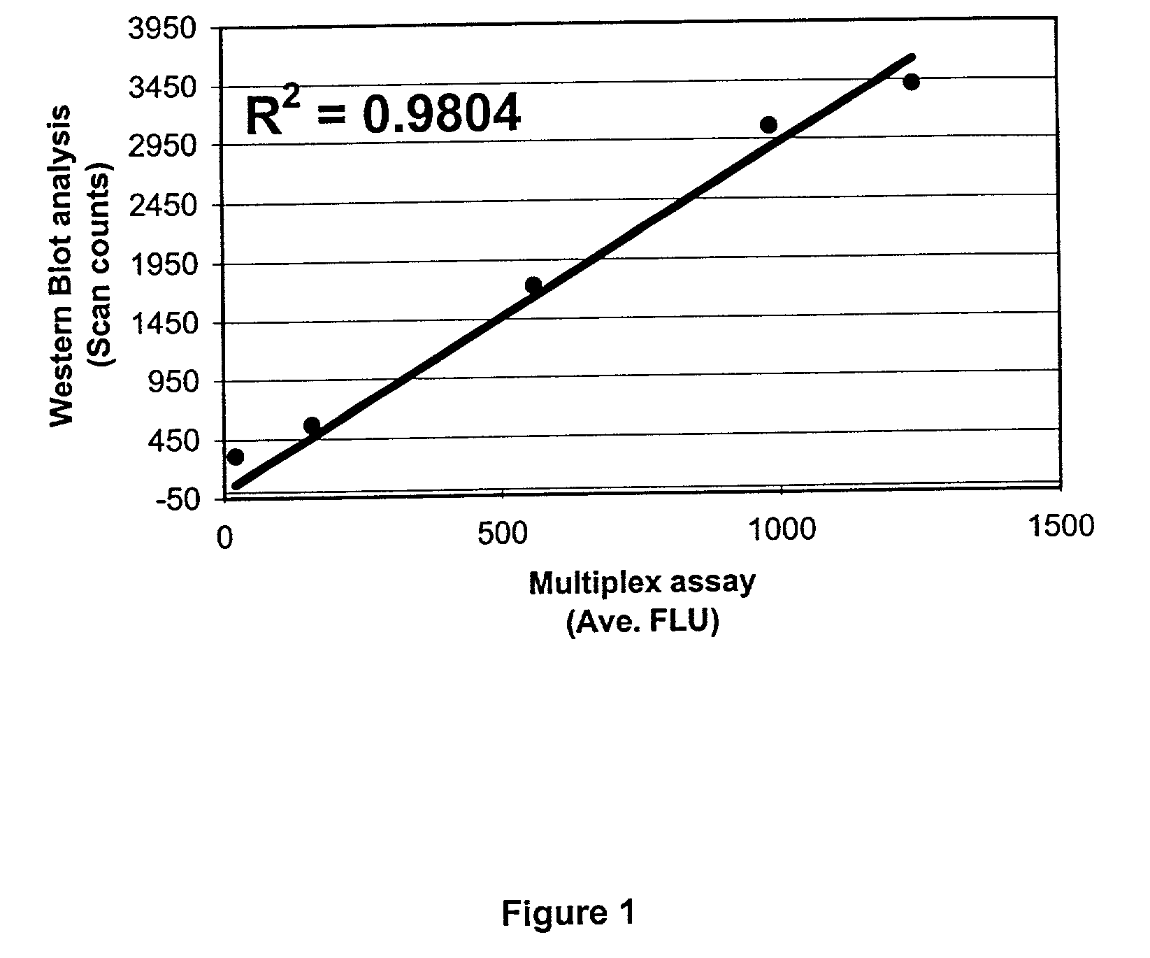
[0093]The above procedure was used to simultaneously analyze a sample of cell lysates containing five phosphorylated proteins: phosphorylated p38MAPK, IκBα, Erk2, JNK and Akt1. The proteins were phosphorylated at the following sites: p38MAPK-Thr180, Tyr182; IκBα-Ser32, Ser36; Erk2-Thr202, Tyr204; JNK-Thr183, Tyr185; Akt1-Ser473. Cell lysates were HeLa and HEK-293 simulated with UV, EGF, Fetal Bovine Serum (FBS) or TNF-alpha. The uninduced HeLa cell lysate was also prepared and used as a negative control. All procedures were done on a 96-well filter plate. The cell lysate samples were analyzed by the above procedure and verified for expression of phosphorylated proteins via Western blotting. The result demonstrates: (1) a simultaneous detection of all five phosphorylated proteins above; (2) only 5–10 μg of cell lysate per well was needed for the analysis; (3) an inter-plate and intra-plate coefficient of variation less than 10% and (4) a tight quan...


PUM

PropertyMeasurementUnit
temperatureaaaaaaaaaa

Abstract

A method and kit for simultaneous detection and / or determination of a plurality of modified proteins in a sample. The method comprises: a) contacting the sample under mild protein denaturation conditions with a plurality of first antibodies capable of binding to a specific target protein, the first antibodies being immobilized on solid support material, each first antibody being differentiable from others by a differentiation parameter, whereby the first antibodies bind to respective target proteins present in the sample; b) removing unbound materials from the locus of the first antibodies; c) contacting the materials from step (b) with one or more second antibodies, each of which is specific to a class or subclass of modified proteins or with a plurality of second antibodies, each of which is specific to a modified protein, so as to bind the second antibody or antibodies to modified proteins in the sample; and d) detecting and / or determining a plurality of modified proteins in the sample. The kit comprises a plurality of primary antibodies immobilized on solid support material, one or more buffers for lysing and for washing cellular material samples to be assayed, an assay buffer for conducting the assay, the buffer containing a sulfate or sulfonate detergent, and one or more second antibodies specific to the modified proteins.

Description

CROSS-REFERENCE TO RELATED APPLICATIONS[0001]This application claims priority of U.S. provisional application No. 60 / 274,130 filed Mar. 7, 2001.BACKGROUND AND PRIOR ART[0002]This invention relates to improvements in processes for conducting “sandwich” assays of proteins and, in particular to an improved method for simultaneously detecting and / or determining, a plurality of modified proteins, especially modified cellular proteins, in a sample. In a typical “sandwich” assay for proteins, a sample (generally a biological fluid or cell extract) comprising a mixture of materials, including one or more proteins, is contacted with a series of microparticles or beads associated with antibodies that bind to the respective target proteins. Each combination of microparticles with a particular antibody has a feature, for instance color, that can distinguish it from the others. After washing away unbound materials, the microparticles, now containing proteins bound to the antibodies, are contacte...

Claims


Application Information

Patent Timeline
Patent Type & Authority Patents(United States)
IPC IPC(8): G01N33/543G01N21/64G01N21/78G01N33/53G01N33/545G01N33/552G01N33/68
CPCG01N33/6803G01N33/6854G01N33/6842Y10S435/973
Inventor NGUYEN, QUANSONG, YONG
Owner BIO RAD LAB INC
Why Eureka
Try Eureka