Endoscopic mucous membrane resection instrument and endoscopic mucous membrane resection method

Active Publication Date: 2006-06-20
OLYMPUS CORP
View PDF9 Cites 55 Cited by
  • Summary
  • Abstract
  • Description
  • Claims
  • Application Information

AI Technical Summary

Benefits of technology

[0019]The present invention may provide an endoscopic mucous membrane resection method included, a resection instrument setting step of fitting an endoscopic mucous membrane resection instrument on a distal end portion of an insertion section of an endoscope, the endoscopic mucous membrane resection instrument including a transparent cap section detachably attached to the distal end portion of the endoscope, one diathermic snare and one ligator, the endoscopic mucous membrane resection instrument being set in a state in which a loop portion of the ligator is engaged in advance on a first projection portion formed at a distal end portion of the cap section and the loop portion is broadened along an inner peripheral surface of the cap section, and also set in a state in which a loop portion of the diathermic snare is engaged on a second projection portion projecting inward at a position spaced apart from the first projection portion and the loop portion is broadened along the inner peripheral surface of the cap section, a step of inserting the endoscope and the resection instrument into a body cavity and moving a distal opening portion of the cap section toward a target to-be-resected mucous membrane, a step of causing a suction force to act within the cap section in a state in which the distal opening portion of the cap section is pushed on the mucous membrane, there

Problems solved by technology

However, in the case of performing endoscopic mucous membrane resection, when a target to-be-resected mucous membran

Method used


Image

  • Endoscopic mucous membrane resection instrument and endoscopic mucous membrane resection method
  • Endoscopic mucous membrane resection instrument and endoscopic mucous membrane resection method
  • Endoscopic mucous membrane resection instrument and endoscopic mucous membrane resection method

Examples


first embodiment

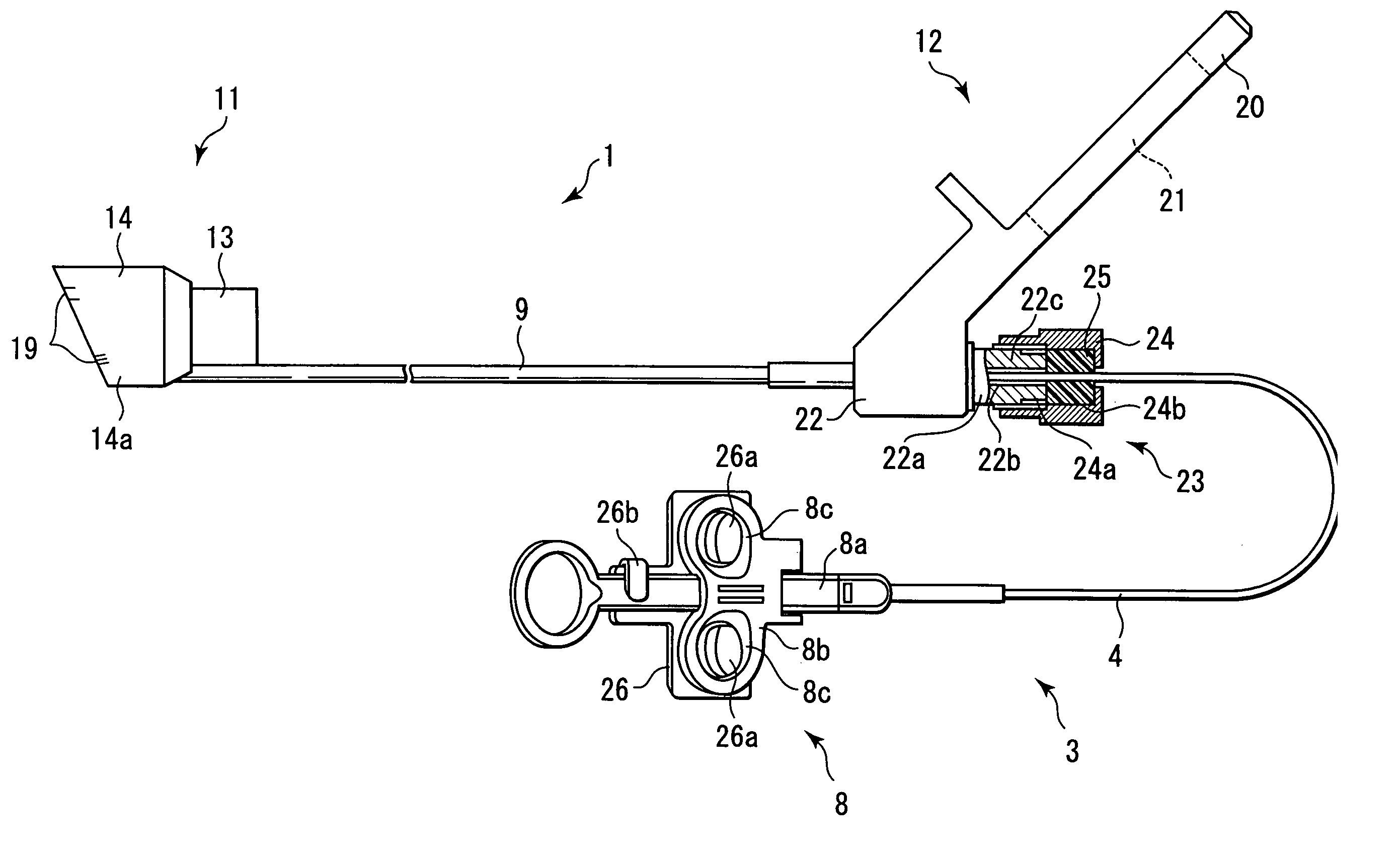
[0086]the present invention will now be described with reference to FIGS. 1A through 12B. FIG. 1A shows a mucous membrane resection instrument 1 for use in an endoscope (hereinafter referred to as “endoscopic mucous membrane resection instrument 1”) according to the embodiment of the invention. As is shown in FIGS. 4A and 4B, the resection instrument 1 is used in combination with an endoscope 2 and two diathermic (high-frequency) snares 3 and 31 serving as endoscopic treatment instruments. The two diathermic snares 3 and 31 have the same structure.

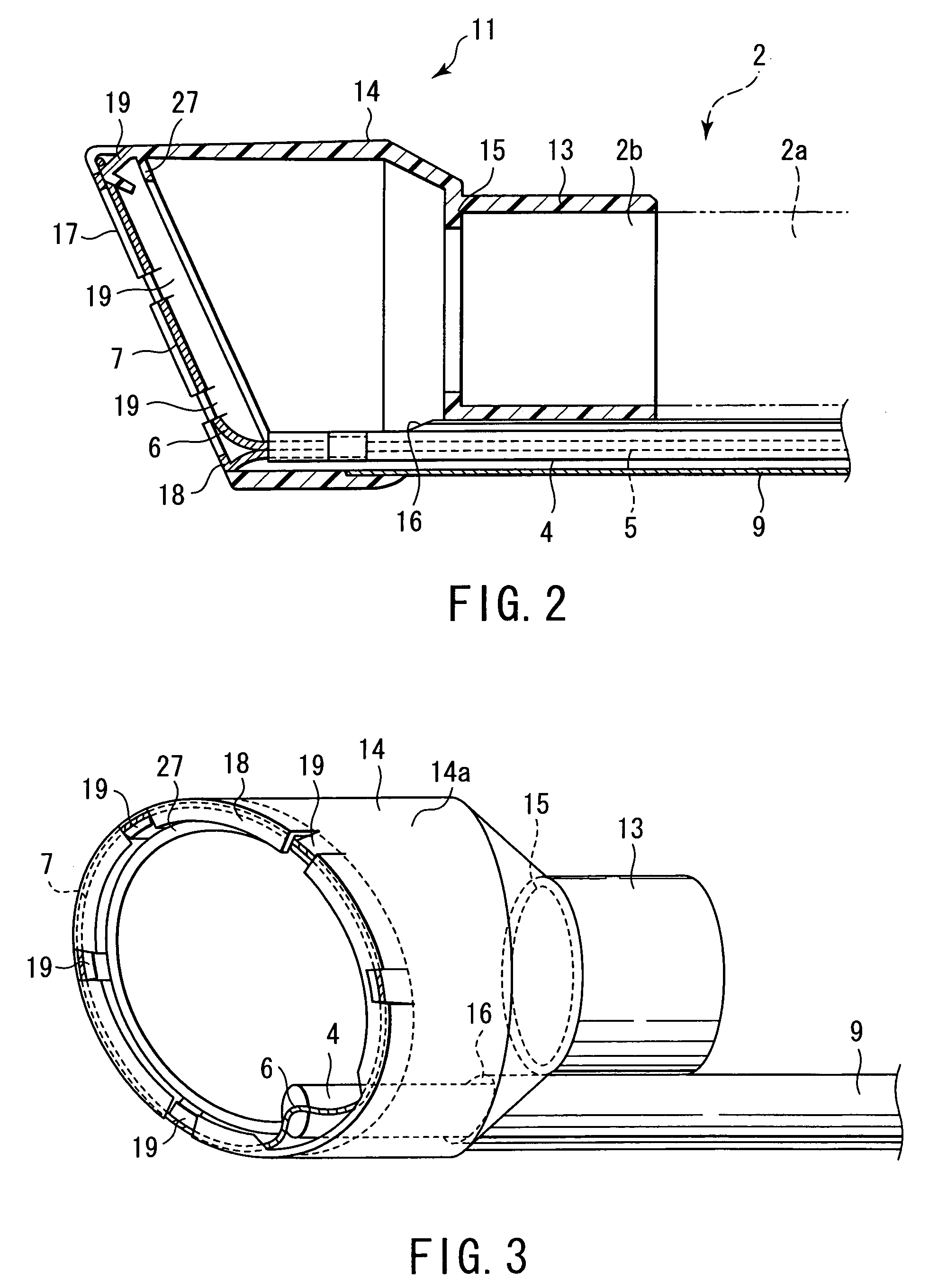
[0087]The endoscope 2 is provided with an elongated insertion section 2a to be inserted in the body. The insertion section 2a has a distal end portion 2b at its distal end. The distal end portion 2b is equipped with an observation optical system, an illumination optical system and a distal opening portion of a treatment instrument insertion channel, although these elements are not shown. Further, a proximal end portion of the insertion sec...

second embodiment

[0125]The endoscopic mucous membrane resection instrument 41 is provided with two tubes 9A and 9B. As is shown in FIG. 15A, two communication port sections 16A and 16B, which communicate with the inside of the cap section 14, are formed at a flange-like stepped portion between the proximal end portion of the cap section 14 of distal-side coupling section 11 and the distal end portion of the endoscope attachment section 13. The two communication port sections 16A and 16B are laterally disposed along the periphery of the cap section 14 at a position where the degree of projection of the inclined face 17 formed at the distal end edge of the cap section 14 is minimum (i.e. at a rearmost position). Further, as shown in FIG. 16A, two tubes 9A and 9B are juxtaposed on the outside of the endoscope attachment section 13. Distal end portions of the tubes 9A and 9B are connected to the communication port sections 16A and 16B.

[0126]The first diathermic snare 3A is inserted in the tube 9A, and ...


PUM


Abstract

One of a first endoscopic treatment instrument setting state and a second endoscopic treatment instrument setting state is selectable. In the former state, when a first diathermic snare is removably inserted in a flexible tube, a loop portion is circumferentially broadened and disposed along a first projection portion. In the latter state, when a second diathermic snare is removably inserted in the flexible tube, a loop portion is circumferentially broadened and disposed along a second projection portion.

Description

CROSS-REFERENCE TO RELATED APPLICATIONS[0001]This application is based upon and claims the benefit of priority from the prior Japanese Patent Application No. 2003-024896, filed Jan. 31, 2003, the entire contents of which are incorporated herein by reference.BACKGROUND OF THE INVENTION[0002]1. Field of the Invention[0003]The present invention relates to a mucous membrane resection instrument for use in an endoscope (hereinafter referred to as “endoscopic mucous membrane resection instrument”), which sucks a mucous membrane in a substantially cylindrical cap attached to a distal end of an insertion portion of an endoscope, thereby forming a polyp-like projection portion, and resects a proximal portion of the projection portion using a diathermic snare, and to a method for the mucous membrane resection.[0004]2. Description of the Related Art[0005]In recent years, endoscopic mucous membrane resection for resecting a mucous membrane of a target part is performed using an endoscope withou...

Claims


Application Information

Patent Timeline
IPC IPC(8): A61B1/04A61B1/00A61B1/012A61B17/30A61B17/32A61B18/14
CPCA61B1/012A61B17/32056A61B18/14A61B2017/00296A61B2017/306A61B2018/1407A61B2017/00269
Inventor OKADA, TSUTOMU
Owner OLYMPUS CORP
Why Eureka
Try Eureka