Capsulated medical equipment

a medical equipment and capsulated technology, applied in the field of capsulated medical equipment, can solve the problems of countermeasures that permit early detection of the clogged state of the capsulated body

Inactive Publication Date: 2006-07-11
OLYMPUS CORP
View PDF12 Cites 39 Cited by
  • Summary
  • Abstract
  • Description
  • Claims
  • Application Information

AI Technical Summary

Benefits of technology

[0010]An object of the present invention is to provide capsulated medical equipment making it possible to detect whether a capsulated body is clogged at a stenosed region in a body cavity.
[0011]Another object of the present invention is to provide capsulated medical equipment making it possible to take appropriate measures in an early stage in case a capsulated body is clogged at a stenosed region in a body cavity.

Problems solved by technology

However, the related art described in the foregoing publication has not disclosed a countermeasure permitting early-stage detection of the clogged state of the capsulated body.

Method used


Image

  • Capsulated medical equipment
  • Capsulated medical equipment
  • Capsulated medical equipment

Examples


first embodiment

[0036](First Embodiment)

[0037]Referring to FIG. 1A to FIG. 9, the first embodiment of the present invention will be described below.

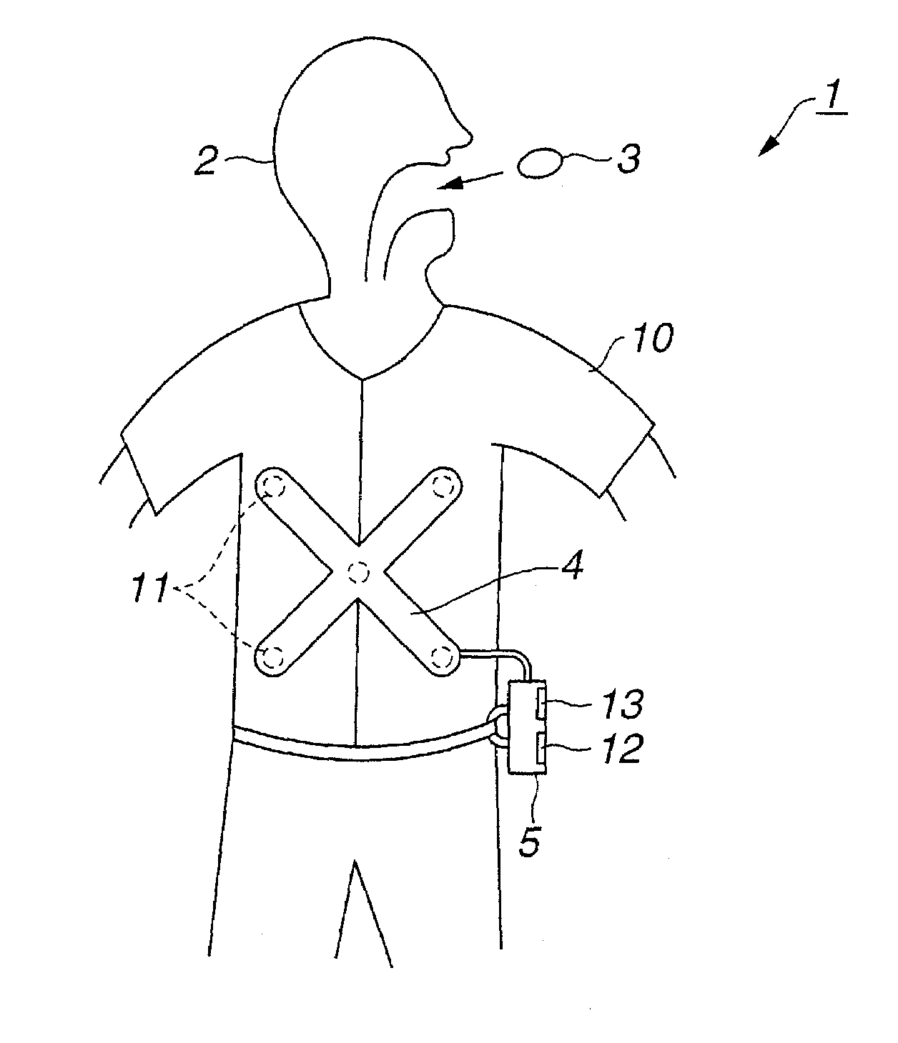
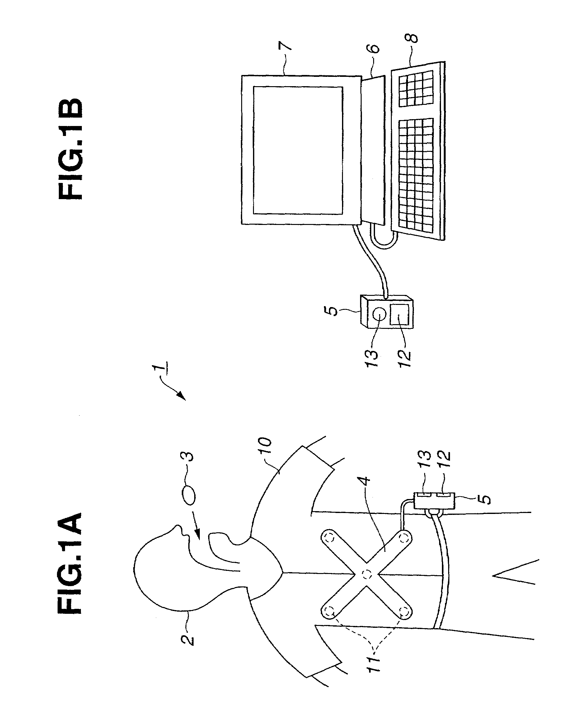
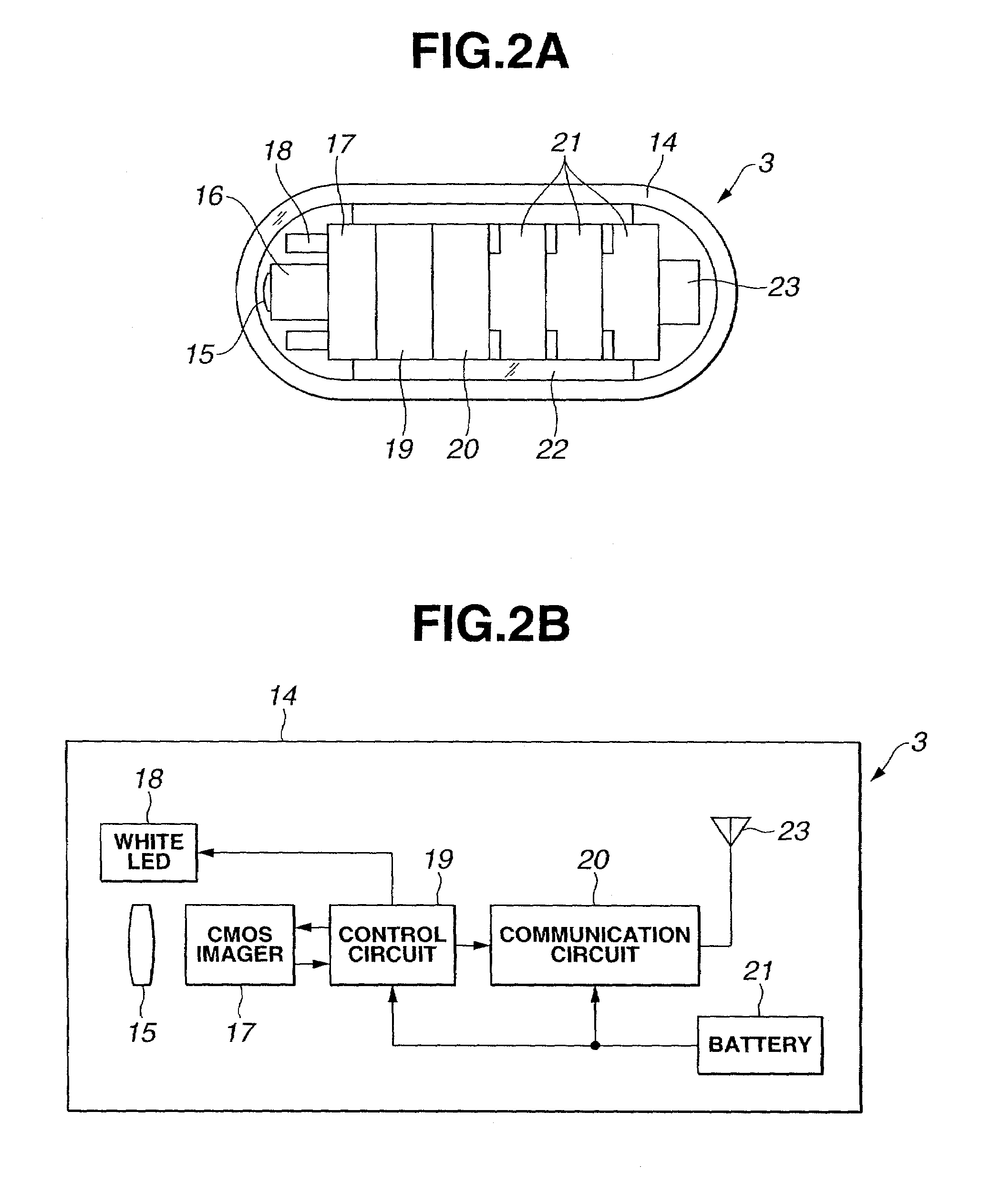
[0038]As shown in FIG. 1A, a capsulated endoscope system 1 in accordance with the first embodiment of the present invention consists mainly of a capsulated endoscope 3 (that is a capsulated body) and an extracorporeal unit 5 (placed away from the patient 2). The capsulated endoscope 3 is gulped down through the mouth of a patient 2, and transmits by radio an image signal that represents an optical image of the inner wall of an intracavitary duct while passing through the intracavitary duct. The extracorporeal unit 5 receives the signal sent from the capsulated endoscope 3 through an antenna unit 4 mounted on the extracorporeal region of the patient 2, and has a facility that preserves images.

[0039]As shown in FIG. 1B, the extracorporeal unit 5 is connected to a personal computer 6 so that it can be disconnected freely. The personal computer 6 fetches an...

second embodiment

[0081](Second Embodiment)

[0082]Next, the second embodiment of the present invention will be described with reference to FIG. 10 to FIG. 12. An object of the present embodiment is to provide capsulated medical equipment having a feature of detecting clogging of a capsulated body (a capsulated endoscope 3C in the present embodiment) in an early stage, and of automatically unclogging the capsulated body. FIG. 10 shows the capsulated endoscope 3C employed in the second embodiment.

[0083]The capsulated endoscope 3C is different from the capsulated endoscope 3 employed in the first embodiment in a point that the capsulated endoscope 3C has a plurality of pressure sensors 43 located on the periphery of a portion thereof having the largest diameter. Moreover, (a stator) of a pager motor (vibrating motor) 44 that is compact and vibrates is locked in the capsulated endoscope at the opposite end of the capsulated endoscope relative to the objective 15. The motor 44 is driven by a motor driver 4...

third embodiment

[0101](Third Embodiment)

[0102]Next, the third embodiment of the present invention will be described with reference to FIG. 13 and FIG. 14. FIG. 13 shows a capsulated endoscope system 51 in accordance with the third embodiment. The system 51 consists mainly of a capsulated endoscope 3D, an extracorporeal unit 52, a personal computer 53, an actuator control circuit 54, actuators 55a and 55b, and electromagnets 56a and 56b. The extracorporeal unit 52 preserves image data produced by the capsulated endoscope 3D and calculates the position of the capsulated endoscope 3D. The personal computer 53 is connected to the extracorporeal unit 52. The actuator control circuit 54 is connected to the personal computer 53. The actuators 55a and 55b are driven with a driving signal sent from the actuator control circuit 54. The electromagnets 56a and 56b are three-dimensionally moved with thrusts produced by the actuators 55a and 55b respectively.

[0103]The capsulated endoscope 3D has the components t...


PUM


Abstract

A capsulated body having a facility, which acquires biomedical information through endoscopic examination or the like, incorporated therein is inserted into a duct within a living body. Whether the capsulated body has halted for a certain period of time is detected from the time-varying position of the capsulated body or a time-passing change in an image. If it is judged that the capsulated body has halted at a stenosed region or the like, the fact is notified so that the capsulated body can be collected immediately.

Description

[0001]The present application claims the benefits of Japanese Application No. 2001-318436 which was filed on Oct. 16, 2001 and the contents of which are incorporated by reference.BACKGROUND OF THE INVENTION[0002]1. Field of the Invention[0003]The present invention relates to capsulated medical equipment that is shaped like a capsule and that moves within a body cavity to perform examination or the like.[0004]2. Description of the Related Art[0005]In recent years, endoscopes and other medical equipment have been proposed for examining an intracavitary region or the like. Moreover, ordinary endoscopes are such that an insertion unit alone is inserted into a body cavity in order to perform endoscopic examination or the like. Medical equipment including a capsulated body that is shaped like a capsule and inserted into a body cavity in order to perform examination or the like has been disclosed in, for example, Japanese Unexamined Patent Application Publication No. 2001-91860.[0006]In th...

Claims


Application Information

Patent Timeline
Patent Type & Authority Patents(United States)
IPC IPC(8): A61B3/16G02B23/24A61B1/00A61B1/04A61B1/273A61B5/00A61B5/07A61B8/13H04N5/225H04N7/18
CPCA61B1/00016A61B1/00156A61B1/00158A61B1/041A61B1/042A61B5/065A61B5/14539A61B5/4839A61B1/273A61B8/565A61B5/0031A61B8/13
Inventor SEGAWA, HIDETAKETAKIZAWA, HIRONOBUADACHI, HIDEYUKIYOKOI, TAKESHI
Owner OLYMPUS CORP
Why Eureka
Try Eureka