Endoscopic submucosal core biopsy device

a biopsy device and submucosal core technology, applied in the field of surgical instruments, can solve the problems of inability to retrieve full tissue core samples, inconvenient design and manufacture, and inability to provide versatility in sampling methods

Inactive Publication Date: 2006-11-21
BOSTON SCI SCIMED INC
View PDF36 Cites 50 Cited by
  • Summary
  • Abstract
  • Description
  • Claims
  • Application Information

AI Technical Summary

Benefits of technology

[0012]It is therefore an object of the invention to provide a surgical instrument that is able to obtain a full core biopsy tissue sample with minimal trauma to the patient.

Problems solved by technology

Many current biopsy instruments, however, cannot retrieve full core samples of tissue, do not provide versatility in sampling methods, or are overly intricate in design and manufacture.
Many current biopsy devices can only take tissue from the surface mucosal layer and also cannot retrieve full core samples.
Further, many current devices lack versatility.
Such practice is time consuming and leads to procedural inefficiency.
Many current instruments also have an overly complex design.
Devices that might otherwise fulfill one of the aforementioned needs, such as the need to capture core samples, for example, often employ complex cutting mechanisms held in sheaths or housings requiring very exact tolerances.

Method used


Image

  • Endoscopic submucosal core biopsy device
  • Endoscopic submucosal core biopsy device
  • Endoscopic submucosal core biopsy device

Examples


Embodiment Construction

[0081]Reference will now be made in detail to the present preferred embodiments of the invention, examples of which are illustrated in the accompanying drawings. Wherever possible the same reference numbers are used in the drawings and the description to refer to the same or like parts.

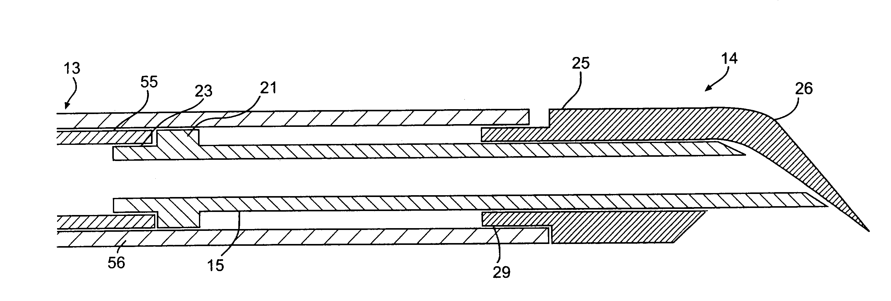
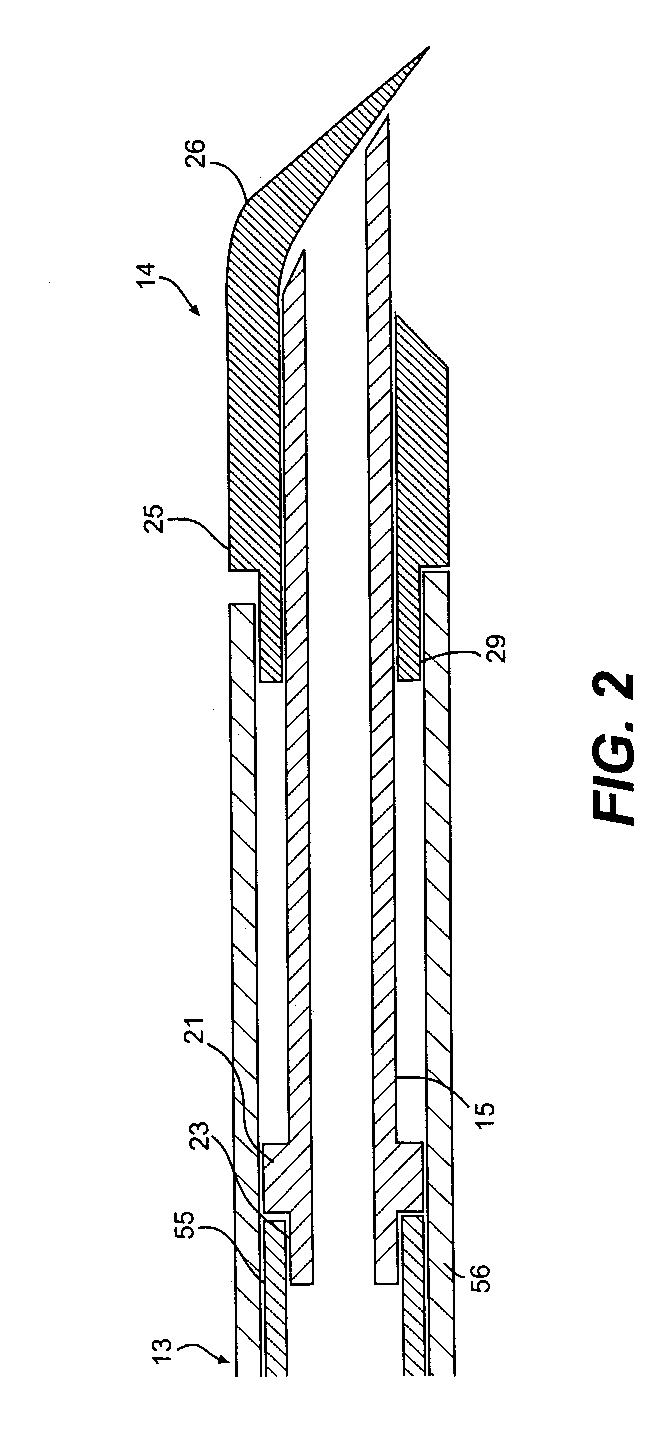
[0082]The present invention is directed to a surgical instrument, particularly an endoscopic instrument for obtaining a biopsy of submucosal tissue. The instrument described in detail below uses a unique end effector to obtain full core biopsy samples and integrates multiple modalities of biopsy tissue sampling. The instrument integrates aspiration and brush cytology with full core biopsy sampling.

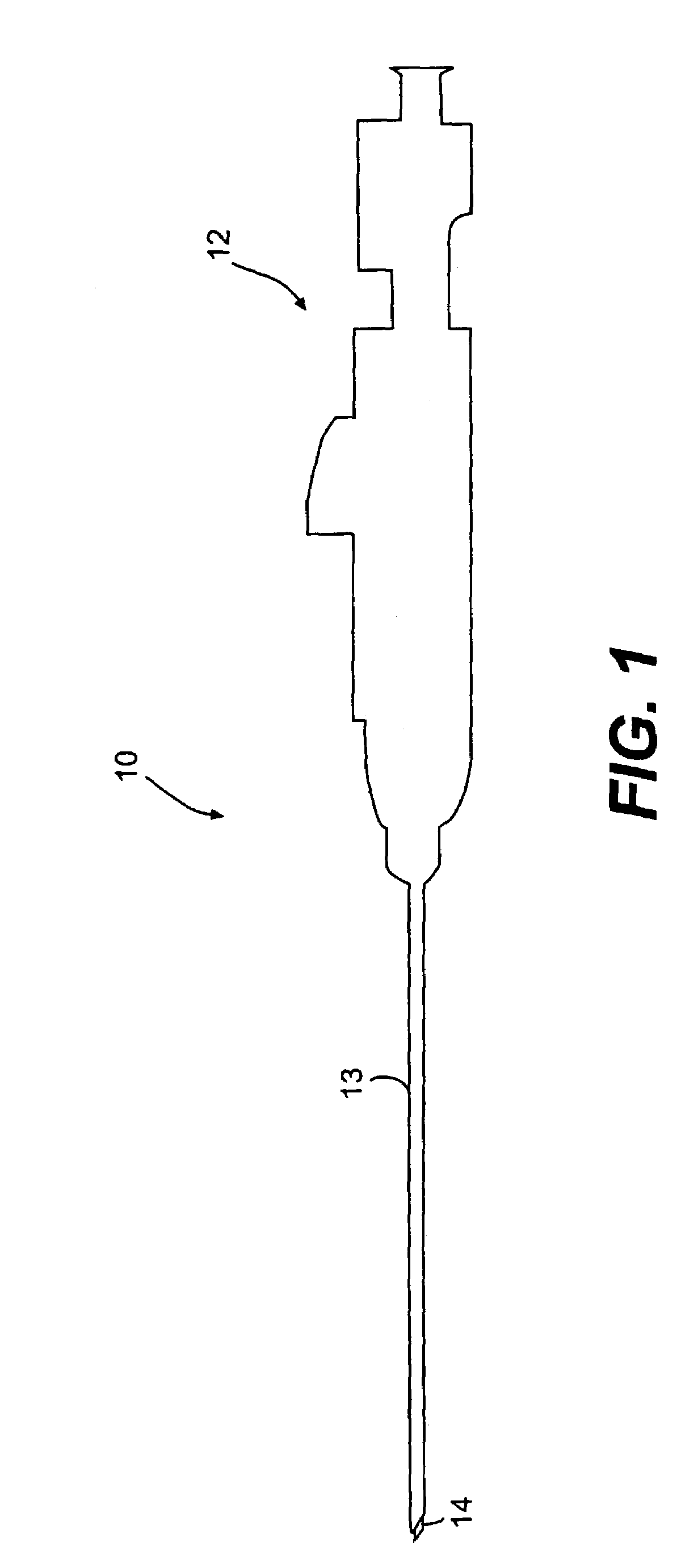
[0083]The instrument according to the present invention is shown generally at 10 in FIG. 1. Instrument 10 includes three main sections: a handle assembly 12 at its proximal end; an end effector assembly 14 at its distal end; and a tubular section 13 extending between handle 12 and end effector assembly 14. E...


PUM


Abstract

An embodiment of the invention includes a device for collecting body tissue. The device includes an inner end effector comprising a hollow portion, a distal edge that defines an opening of the hollow portion at least a portion of which is sufficiently sharpened to cut body tissue, and a proximal end configured to receive an inner tubular member. The device also includes an outer end effector comprising a flexible extension on a distal end with an edge at least a portion of which is sufficiently sharpened to cut body tissue and biased inwardly toward the hollow portion of the inner end effector to cover the opening, and a proximal end configured to receive an outer tubular member. The inner end effector is slidably disposed within the outer end effector.

Description

[0001]This is a continuation of U.S. patent application Ser. No. 09 / 801,746 filed Mar. 9, 2001, now U.S. Pat. No. 6,551,254, which is a continuation of U.S. patent application Ser. No. 09 / 406,917 filed Sep. 28, 1999, now U.S. Pat. No. 6,248,081, both of which are incorporated herein by reference.BACKGROUND OF THE INVENTION[0002]1. Field of the Invention[0003]The present invention relates to surgical instruments and, particularly, to an instrument for biopsy sampling of tissue. Still more particularly, the present invention relates to an endoscopic biopsy instrument that obtains core biopsy samples and integrates two or more biopsy-sampling modalities.[0004]2. Background of Related Art[0005]Biopsy is the removal and study of body tissue for medical diagnosis. Typically, physicians obtain biopsy samples in order to detect abnormalities such as cancer and determine the extent to which cancerous tissue has spread. They use various biopsy instruments to acquire tissue samples from differ...

Claims


Application Information

Patent Timeline
Patent Type & Authority Patents(United States)
IPC IPC(8): A61B10/00A61B10/02A61B10/04
CPCA61B10/0266A61B10/04A61B17/32053A61B2010/0216A61B2017/00269A61B2017/32004
Inventor NISHTALAS, SRINIVASWARD, TIM E.BATES, JIMGOBRON, STEPHANE
Owner BOSTON SCI SCIMED INC
Why Eureka
Try Eureka