Enhanced X-ray imaging system and method

a x-ray imaging and enhanced technology, applied in the field of non-invasive imaging, can solve the problems of difficult interpretation of single-view x-ray images associated with mammography, inability to detect attenuation differences or structural distortions, and inability to provide as much information or detail as desired

Active Publication Date: 2006-11-28
GENERAL ELECTRIC CO
View PDF7 Cites 224 Cited by
  • Summary
  • Abstract
  • Description
  • Claims
  • Application Information

AI Technical Summary

Problems solved by technology

The X-rays are attenuated by the breast tissue, with abnormalities presumably giving rise to discernible attenuation differences or structural distortions, and impact a detector.
While mammography is a useful tool for breast examination and screening, the images acquired by mammography may not provide as much information or detail as desired.
In particular, the single view X-ray images associated with mammography may be difficult to interpret since all of the anatomic structure of the imaged breast is superimposed in the image.
As a result, the sensitivity rate for X-ray mammography is relatively low (typically between 70% and 80%) and the false positive rate is undesirably high (between 70% and 90% of biopsies are normal).

Method used


Image

  • Enhanced X-ray imaging system and method
  • Enhanced X-ray imaging system and method
  • Enhanced X-ray imaging system and method

Examples


example

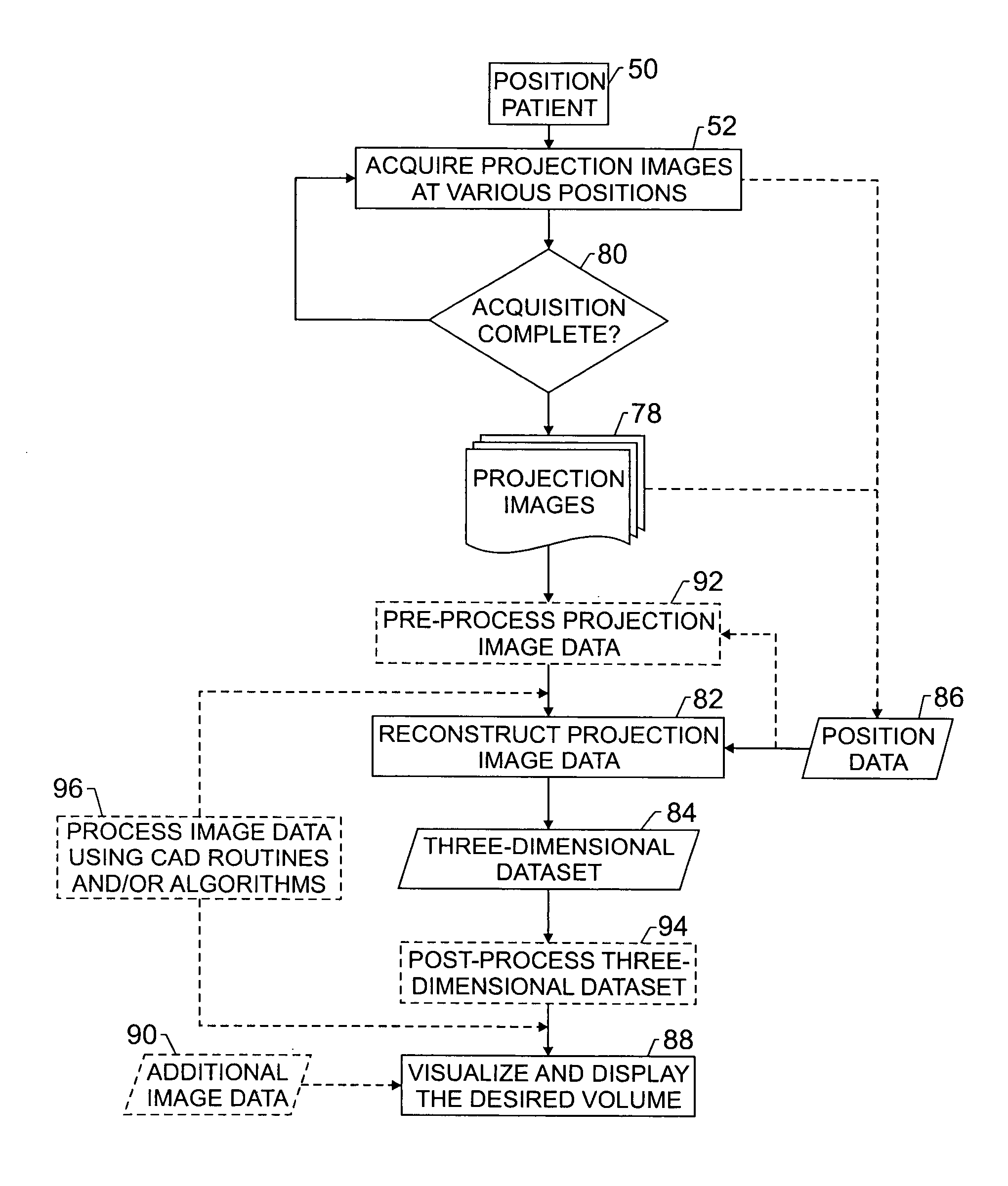
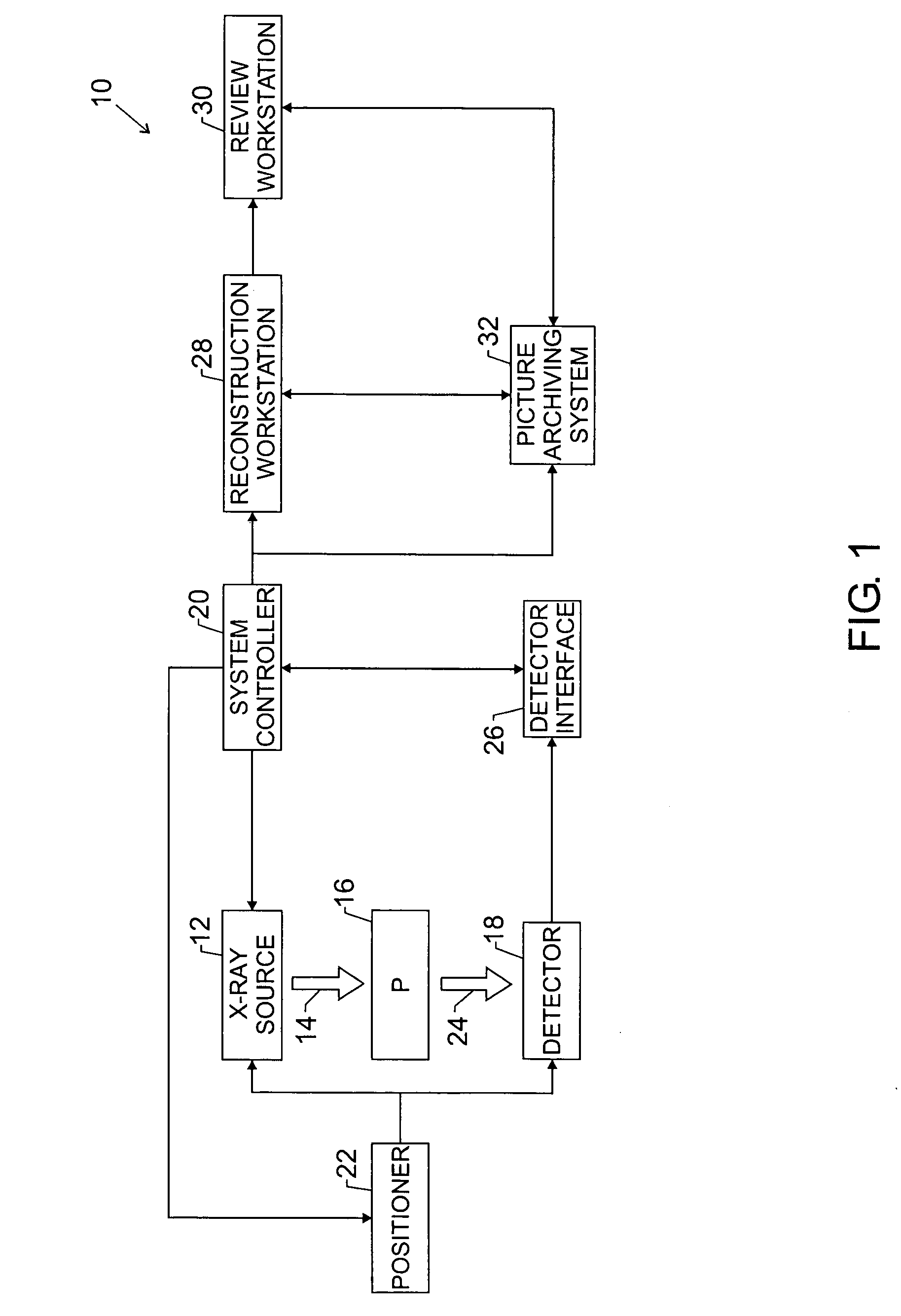
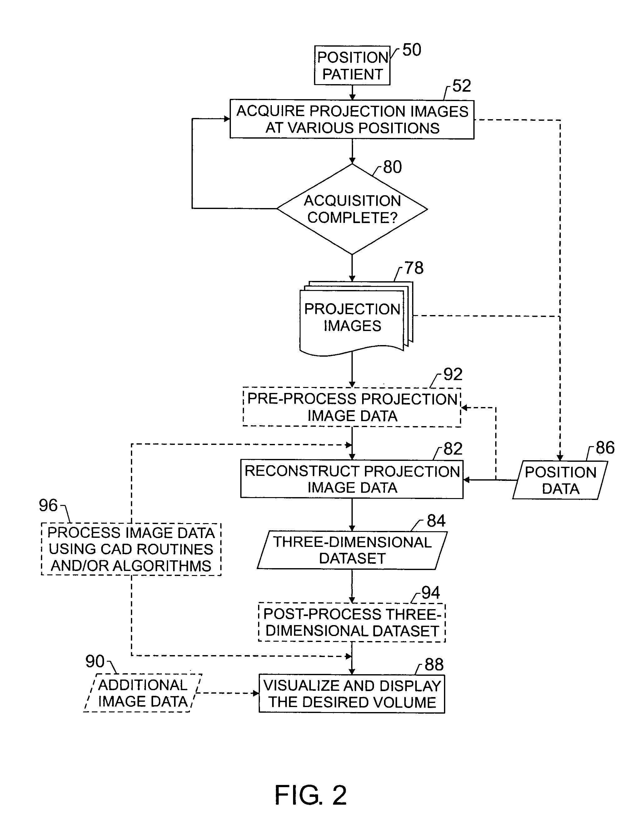
[0078]An exemplary implementation of the foregoing technique is now presented. In this exemplary implementation a three-dimensional imaging system 10 acquires twenty-one projections over a 60° angular range in approximately eight seconds. The X-ray source 12 includes an X-ray tube which moves in a trajectory above the detector 22 and stationary breast. The Source to Image Distance (SID) is 660 mm. For a relatively thick, dense breast (such as 6 cm of compressed thickness), a technique of Rh / Rh at 30 kVp and 160 mAs total may be used. The mAs per tomosynthesis view is 160 / 21=7.62 mAs. The X-ray tube current is approximately 75 mA, for an X-ray “on” time per shot of approximately 0.1 sec. Both the X-ray tube and the detector 22 move during each X-ray exposure. In this example, the X-ray tube moves by approximately 240 microns and the detector 22 moves by approximately 20 microns. The X-ray tube also moves between exposures. Total dose for the tomosynthesis mammogram is approximately 1...


PUM

PropertyMeasurementUnit
inherent depthaaaaaaaaaa
Source to Image Distanceaaaaaaaaaa

Abstract

Techniques are provided for generating three-dimensional images, such as may be used in mammography. In accordance with these techniques, projection images of an object of interest are acquired from different locations, such as by moving an X-ray source along an arbitrary imaging trajectory between emissions or by individually activating different X-ray sources located at different locations relative to the object of interest. The projection images may be reconstructed to generate a three-dimensional dataset representative of the object from which one or more volumes may be selected for visualization and display. Additional processing steps may occur throughout the image chain, such as for pre-processing the projection images or post-processing the three-dimensional dataset.

Description

BACKGROUND OF THE INVENTION[0001]The present invention relates generally to the field of non-invasive imaging and more specifically to the field of medical imaging. In particular, the present invention relates to the generation of three-dimensional image data, such as in a mammography context.[0002]Mammography is an imaging technique by which a breast may be non-invasively examined or screened to detect abnormalities, such as lumps, fibroids, lesions, calcifications, and so forth. Typically mammography employs radiographic techniques to generate images representative of the breast tissue. In particular, the breast is typically compressed to near uniform thickness and X-rays are passed through the compressed breast. The X-rays are attenuated by the breast tissue, with abnormalities presumably giving rise to discernible attenuation differences or structural distortions, and impact a detector. The detector, in turn generates responsive signals which may be processed to generate an imag...

Claims


Application Information

Patent Timeline
Patent Type & Authority Patents(United States)
IPC IPC(8): G01N23/04A61B5/05A61B6/00
CPCA61B6/482A61B6/584A61B6/502A61B6/4007A61B6/4035A61B6/027Y10S378/901
Inventor EBERHARD, JEFFREY WAYNECLAUS, BERNHARD ERICH HERMANNLANDBERG, CYNTHIA
Owner GENERAL ELECTRIC CO
Why Eureka
Try Eureka