
Treatment and diagnosis of cancer

a biological agent and cancer technology, applied in the field of cancer treatment and diagnosis, can solve the problems of inability to distinguish metastatic prostate cancer involvement of lymph nodes by a criterion other than size (i.e. >1 cm), and continue to challenge the medial community, so as to achieve more effective treatment and diagnosis.

Inactive Publication Date: 2007-01-16
CORNELL RES FOUNDATION INC
View PDF98 Cites 94 Cited by
  • Summary
  • Abstract
  • Description
  • Claims
  • Application Information

AI Technical Summary

Benefits of technology

[0037]The biological agents of the present invention recognize the extracellular domain of antigens of normal, benign hyperplastic, and cancerous prostate epithelial cells. Unlike the 7E11 antibody, which recognizes an epitope of prostate-associated antigens which are exposed extracellularly only after cell lysis, the biological agents of the present invention bind to antigenic epitopes which are extracellularly exposed in living prostate cells. Using the biological agents of the present invention, living, unfixed normal, benign hyperplastic, and cancerous prostate epithelial cells can be targeted, which makes treatment and diagnosis more effective. In a preferred embodiment for treating prostate cancer, the biological agents of the present invention also bind to and are internalized with the prostate specific membrane antigen, which permits the therapeutic use of intracellularly acting cytotoxic agents.

Problems solved by technology

In spite of improved treatments for certain forms of cancer, it is still a leading cause of death in the United States.
However, the development of methods that permit rapid and accurate detection of many forms of cancers continues to challenge the medial community.
Accurate clinical evaluation of nodal involvement has proven to be difficult.
Imaging techniques such as computed tomography (“CT”) or magnetic resonance (“MR”) imaging are unable to distinguish metastatic prostate cancer involvement of lymph nodes by criterion other than size (i.e., >1 cm).
Therefore, by definition, these imaging modalities are inherently insensitive in the detection of small volume (<1 cm) disease as well as non-specific in the detection of larger volume adenopathy.
Prior art attempts to develop a specific test for prostatic acid phosphatase have met with only limited success, because techniques which rely on enzyme activity on a so-called “specific” substrate cannot take into account other biochemical and immunochemical differences among the many acid phosphatases which are unrelated to enzyme activity of prostate origin.
In addition to the aforementioned problems of non-specificity which appear to be inherent in many of the prior art reagents employed for the detection of prostate acid phosphatase, there have been reports of elevated serum acid phosphatase associated with other diseases, which further complicates the problem of obtaining an accurate clinical diagnosis of prostatic cancer.
However, the previously reported immunochemical methods have drawbacks of their own which have precluded their widespread acceptance.
However, this method has the disadvantages of limited sensitivity, even with the large amounts of antigen employed, and of employing antisera which may cross-react with other, antigenically unrelated serum protein components present in prostatic fluid.
However, practical problems are posed by the need for a source of cancerous prostate tissue from which the diagnostically relevant prostatic acid phosphatase isoenzyme patterns associated with prostatic cancer are extracted for the preparation of antibodies thereto.
Historically, the drawback of this procedure is that most cancers had spread beyond the bounds of the operation by the time they were detected.
Patients with bulky, high-grade tumors are less likely to be successfully treated by radical prostatectomy.
Diethylstilbestrol from estrogen is another useful hormonal therapy which has a disadvantage of causing cardiovascular toxicity.
As a result, it blocks the effect of testosterone, increasing serum testosterone concentrations and allows patients to remain potent—a significant problem after radical prostatectomy and radiation treatments.
Cytotoxic chemotherapy is largely ineffective in treating prostate cancer.
Its toxicity makes such therapy unsuitable for elderly patients.
While many mAbs have previously been prepared against prostate related antigens, none of these mAbs were specifically generated with an imaging objective in mind.
However, the antigens detected by these monoclonal antibodies are present in the blood and, therefore, compete with antigens at tumor sites for the monoclonal antibodies.
This causes background noise which makes the use of such antibodies inadequate for in vivo imaging.
In therapy, such antibodies, if bound to a cytotoxic agent, could be harmful to other organs.
Consequently, imaging using 7E11 is limited to the detection of dead cells within tumor deposits.
Additionally, the therapeutic use of the 7E11 antibody is limited, because only cells that are already dead or tissue containing a large proportion of dead cells can be effectively targeted.

Method used


Image

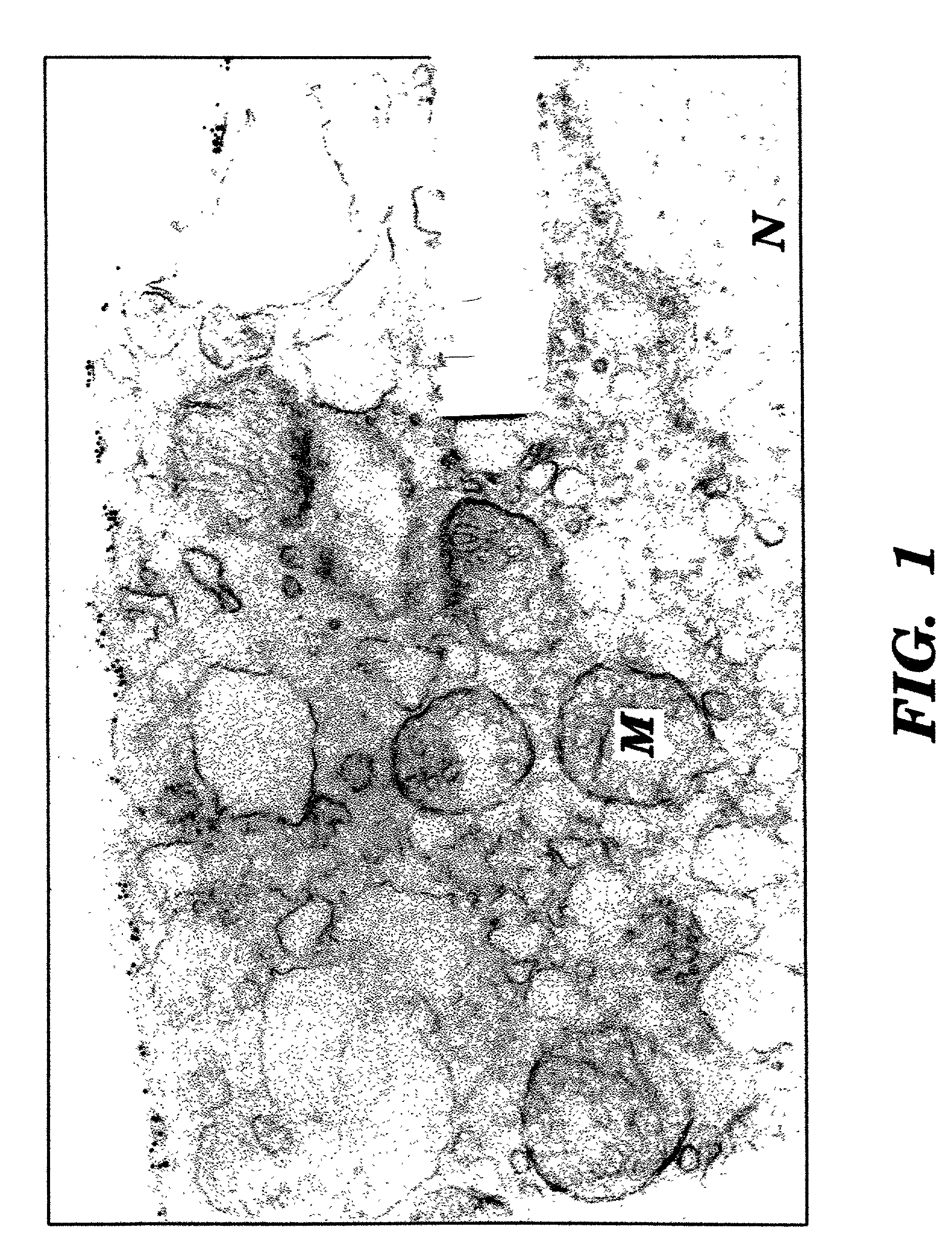
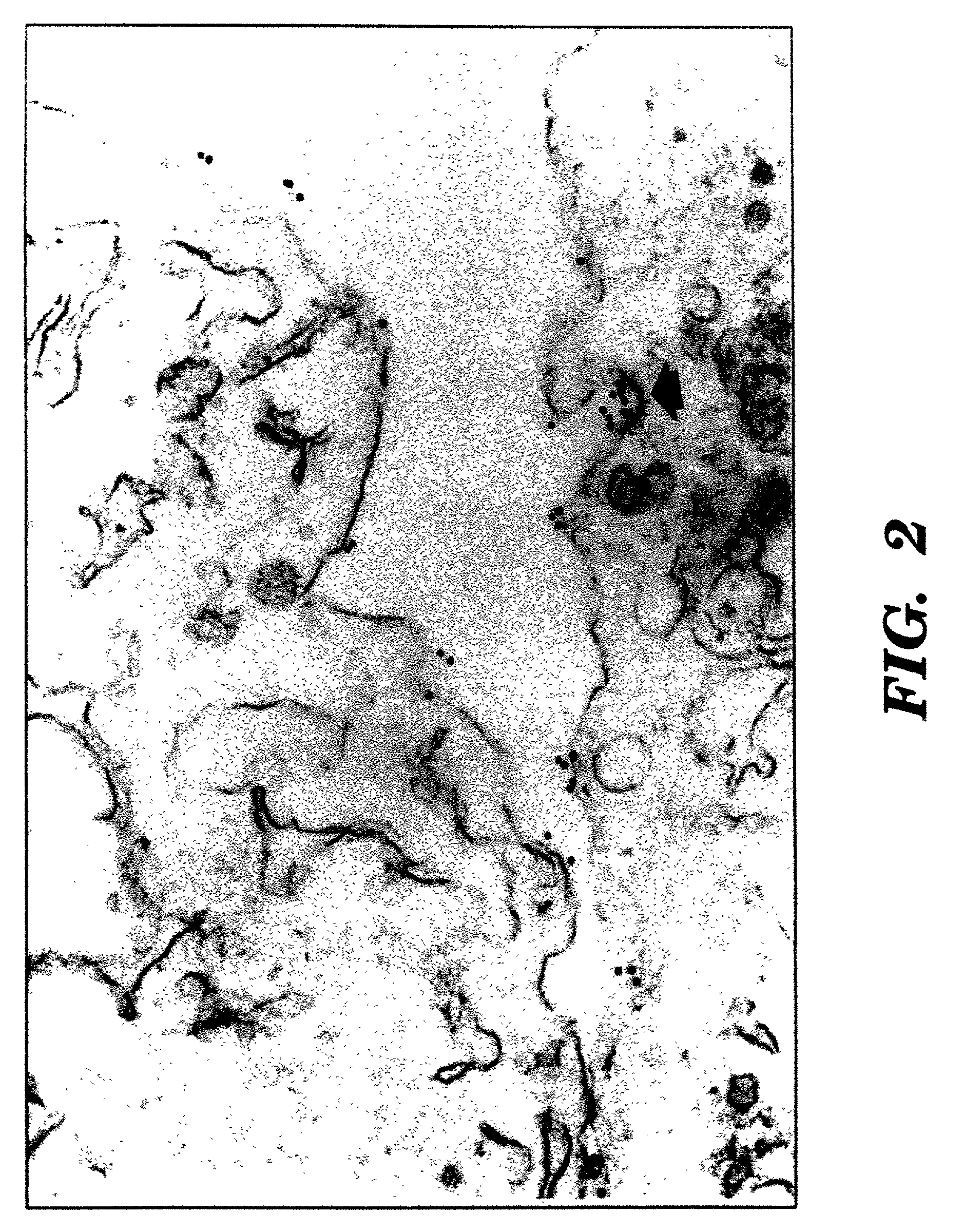
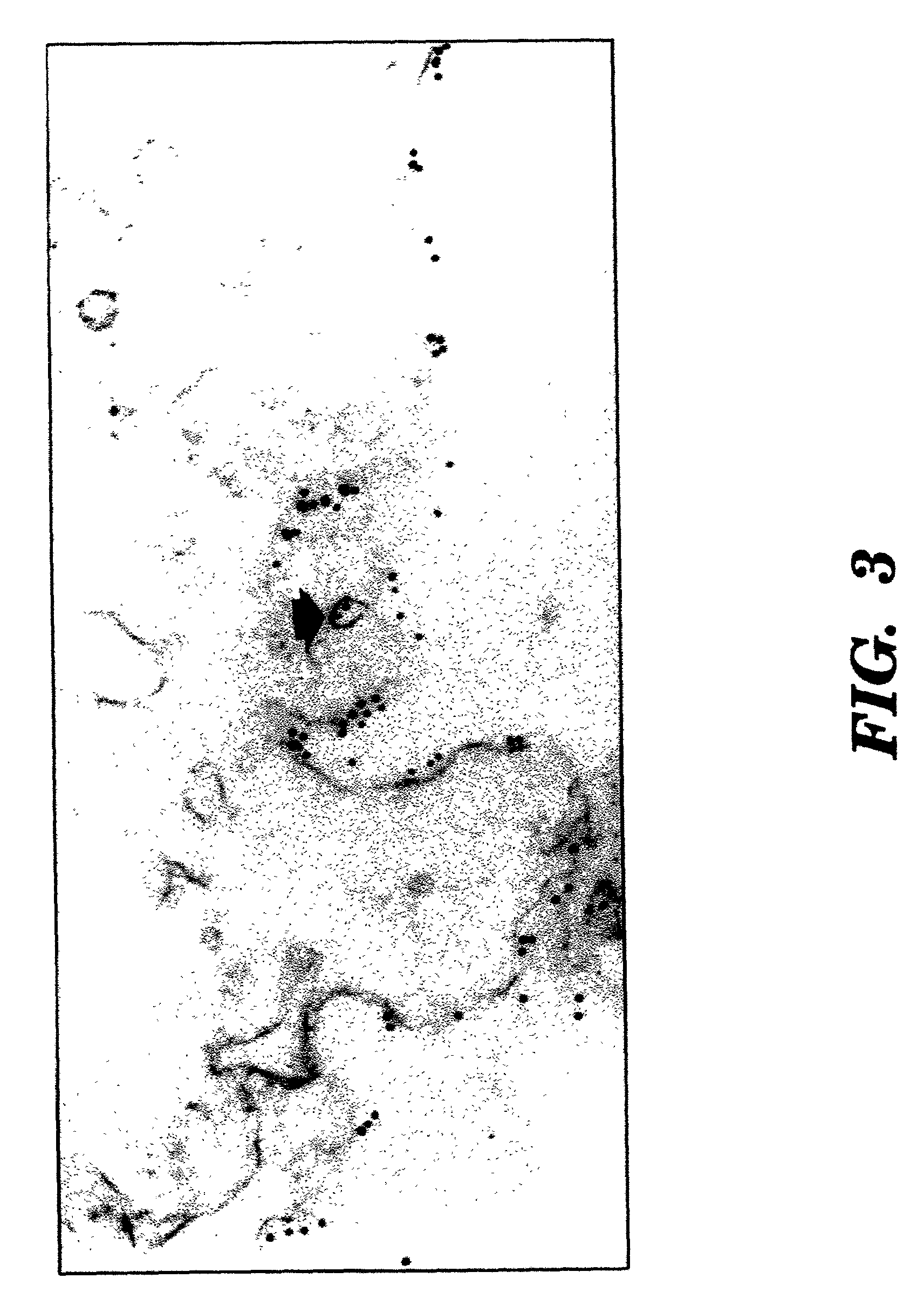
  • Treatment and diagnosis of cancer
  • Treatment and diagnosis of cancer
  • Treatment and diagnosis of cancer

Examples


example 1

Human Tissues

[0099]Fresh specimens of benign and malignant tissues were obtained from the Department of Pathology of New York Hospital Cornell University Medical Center (“NYH-CUMC”).

example 2

Tissue Culture

[0100]Cultured cell lines of human cancers were obtained from the Laboratory of Urological Oncology of NYH-CUMC. The prostate cancer cell lines PC-3 (Mickey, D. D., et al., “Characterization Of A Human Prostate Adenocarcinoma Cell Line (DU145) As A Monolayer Culture And As A Solid Tumor In Athymic Mice,”Prog. Clin. Biol. Res., 37:67–84 (1980), which is hereby incorporated by reference), DU-145 (Mickey, D. D., et al., “Characterization Of A Human Prostate Adenocarcinoma Cell Line (DU145) As A Monolayer Culture And As A Solid Tumor In Athymic Mice,”Prog. Clin. Biol. Res., 37:67–84 (1980), which is hereby incorporated by reference), and LNCaP (Horoszewicz, J. S., et al., “LNCaP Model Of Human Prostatic Carcinoma,”Cancer Res., 43:1809–1818 (1983), which is hereby incorporated by reference) were obtained from the American Type Culture Collection (Rockville, Md.). Hybridomas were initially cloned in RPMI-1640 medium supplemented with 10% FCS, 0.1 mM nonessential amino acids,...

example 3

Preparation of Mouse Monoclonal Antibodies

[0101]Female BALB / c mice were immunized intraperitoneally with LNCaP (6×10⁶ cells) three times at 2 week intervals. A final intraperitoneal booster immunization was administered with fresh prostate epithelial cells which had been grown in vitro. Three days later, spleen cells were fused with SP-2 mouse myeloma cells utilizing standard techniques (Ueda, R., et al., “Cell Surface Antigens Of Human Renal Cancer Defined By Mouse Monoclonal Antibodies: Identification Of Tissue-Specific Kidney Glycoproteins,”Proc. Natl. Acad. Sci. USA, 78:5122–5126 (1981), which is hereby incorporated by reference). Supernatants of the resulting clones were screened by rosette and complement cytotoxicity assays against viable LNCaP. Clones which were positive by these assays were screened by immunochemistry vs normal kidney, colon, and prostate. Clones which were LNCap+ / Nm1Kid− / colon− / prostate+ were selected and subcloned 3 times by limiting dilution. The immunogl...


PUM

PropertyMeasurementUnit
sizeaaaaaaaaaa
wavelengthsaaaaaaaaaa

Abstract

The present invention is directed to the use of antibodies or binding portions thereof, probes, ligands, or other biological agents which either recognize an extracellular domain of prostate specific membrane antigen or bind to and are internalized with prostate specific membrane antigen. These biological agents can be labeled and used for detection of cancerous tissues, particularly cancerous tissues proximate to or containing vascular endothelial cells, which express an extracellular domain of prostate specific membrane antigen. The labeled biological agents can also be used to detect normal, benign hyperplastic, and cancerous prostate epithelial cells or portions thereof. They also can be used alone or bound to a substance effective to ablate or kill such cells as a therapy for prostate or other cancers. Also disclosed are four hybridoma cell lines, each of which produces a monoclonal antibody recognizing extracellular domains of prostate specific membrane antigens of normal, benign hyperplastic, and cancerous prostate epithelial cells or portions thereof.

Description

[0001]This application is a Continuation Application of application Ser. No. 09 / 357,708, filed on Jul. 20, 1999 now U.S. Pat. No. 6,770,450; which is a Divisional of application Ser. No. 08 / 895,914, filed on Jul. 17, 1997 now U.S. Pat. No. 6,136,311; which is a continuation-in-part of U.S. patent application Ser. No. 08 / 838,682, filed Apr. 9, 1997, now U.S. Pat. No. 6,107,090 which claims the benefit of U.S. Provisional Patent Application Ser. No. 60 / 016,976, filed May 6, 1996; and U.S. Provisional Patent Application Ser. No. 60 / 022,125, filed Jul. 18, 1996.FIELD OF THE INVENTION[0002]The present invention relates to the treatment and diagnosis of cancer with biological agents.BACKGROUND OF THE INVENTION[0003]In spite of improved treatments for certain forms of cancer, it is still a leading cause of death in the United States. Since the chance for complete remission of cancer is, in most cases, greatly enhanced by early diagnosis, it is very desirable that physicians be able to dete...

Claims


Application Information

Patent Timeline
Patent Type & Authority Patents(United States)
IPC IPC(8): A61K39/395A61P35/00C07K16/30C12P21/08G01N33/574
CPCA61K47/48638A61K47/48761C07K16/3069G01N33/57434B82Y5/00A61K2039/505G01N2333/705C07K2317/77A61K47/6869A61K47/6899A61P35/00
Inventor BANDER, NEIL H.
Owner CORNELL RES FOUNDATION INC
Learn More