Ultrasonic Imaging Apparatus and Projection Image Generating Method

a technology of ultrasonic imaging and projection image, which is applied in the field of ultrasonic imaging apparatus and projection image generating method, can solve the problems of inconvenient devices and difficult grasping, and achieve the effect of convenient stereoscopic and effective diagnosis and effective diagnosis

Active Publication Date: 2008-10-23
FUJIFILM HEALTHCARE CORP
View PDF5 Cites 39 Cited by
  • Summary
  • Abstract
  • Description
  • Claims
  • Application Information

AI Technical Summary

Benefits of technology

[0010]According to this invention, the first projection image is generated by adding the first three-dimensional image data with the information of the second three-dimensional image data, and thus the information on the overlap with the second three-dimensional image data is reflected to brightness every pixel. Accordingly, by referring to the shading (grayscale) of the first projection image, the overlap condition with the second three-dimensional image data can be grasped, and a stereoscopic and effective diagnosis can be easily performed.

Problems solved by technology

Accordingly, in order to surely grasp the stereoscopic positional relationship between the blood vessel and the tissues around the blood vessel (for example, the penetration state of the blood vessel through the tissues), it is necessary to carry out an operation of rotating the display angle of the composite image or remove unnecessary image data from the composite image, and thus there is a problem that this device is inconvenient.
Furthermore, when the tissue projection image and the Doppler projection image are displayed in superposition with each other while varying the composition rate between the tissue projection image and the Doppler projection image, the brightness of the respective images is equally changed in connection with the change of the composition rate, and thus it is difficult to grasp, from the displayed composite image, the stereoscopic positional relationship between the blood vessel and the tissue around the blood vessel, for example, the overlap condition of the respective images when viewed from the projection direction, depth feel in the projection direction in the overlap area, etc.

Method used


Image

  • Ultrasonic Imaging Apparatus and Projection Image Generating Method
  • Ultrasonic Imaging Apparatus and Projection Image Generating Method
  • Ultrasonic Imaging Apparatus and Projection Image Generating Method

Examples


Embodiment Construction

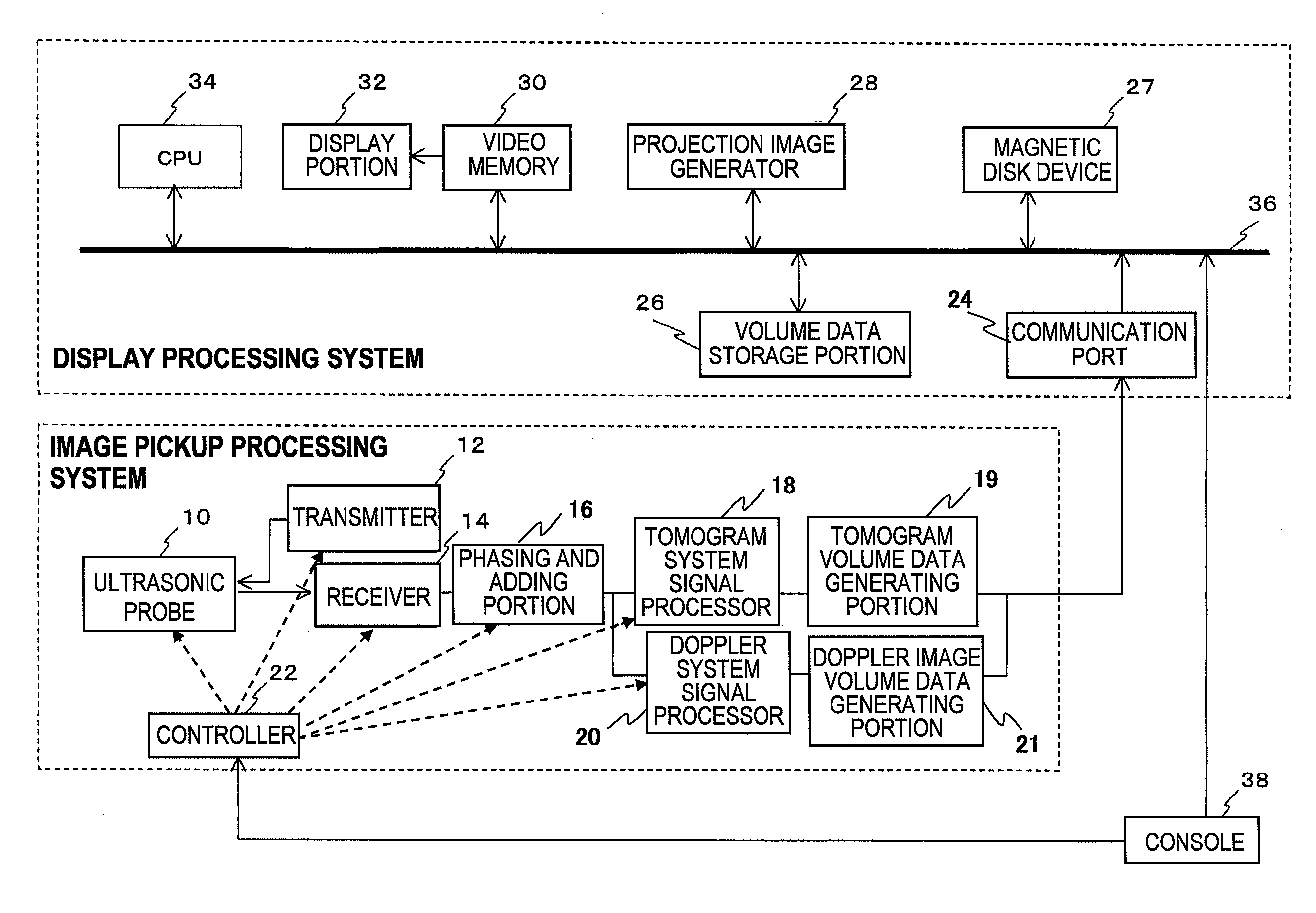
[0021]An embodiment of an ultrasonic imaging apparatus and a projection image generating method to which the present invention is applied will be described with reference to FIGS. 1 to 8. FIG. 1 is a diagram showing the construction of an ultrasonic imaging apparatus of this embodiment.

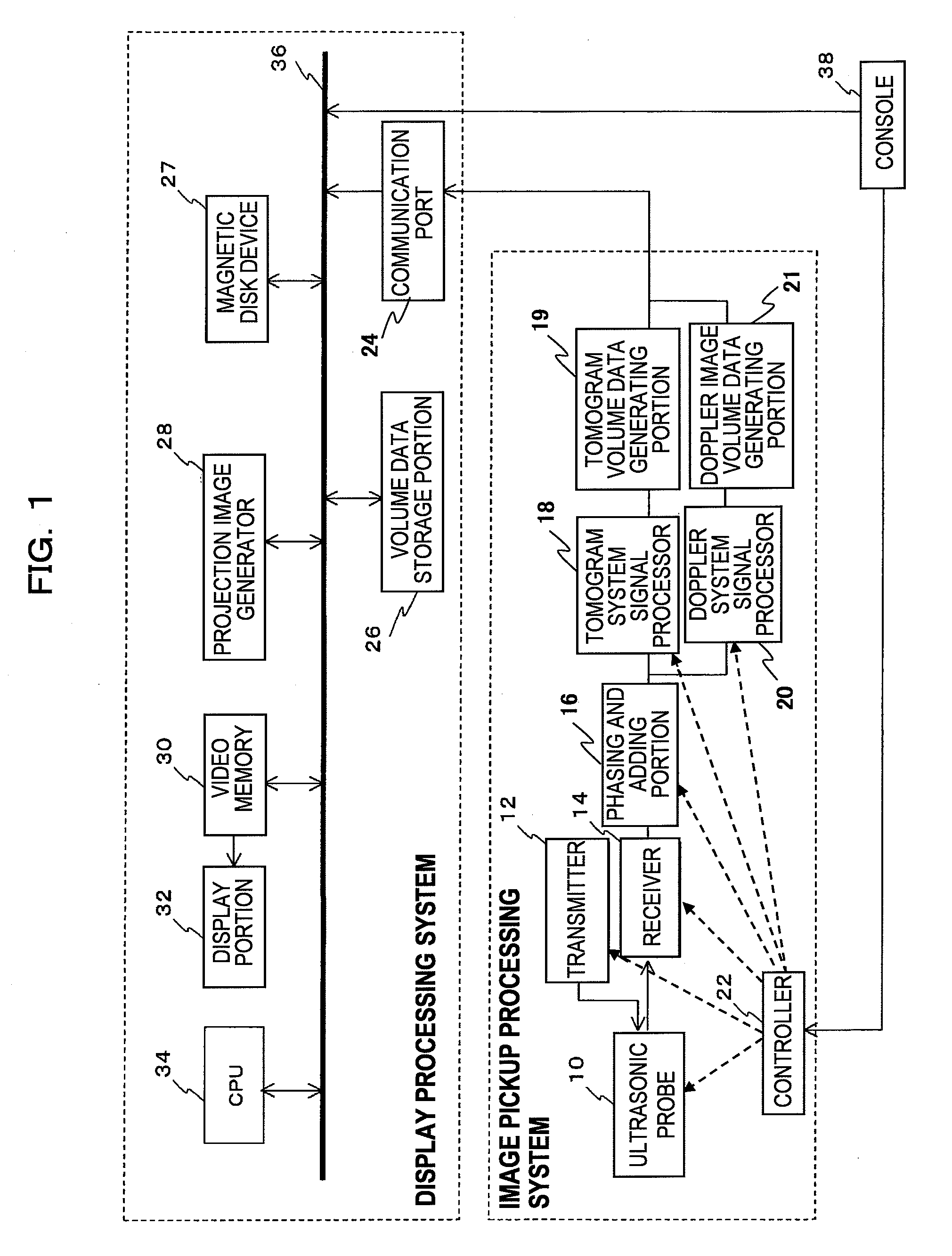
[0022]As shown in FIG. 1, the ultrasonic imaging apparatus is roughly divided into an image pickup processing system of obtaining a three-dimensional ultrasonic image volume data of an object being examined and a display processing system of displaying the obtained three-dimensional ultrasonic image volume data.

[0023]The imaging apparatus is equipped with an ultrasonic probe 10 including plural vibrators that are arranged two-dimensionally and transmit / receive ultrasonic waves to / from an object being examined, a transmitter 12 for supplying a driving signal to the ultrasonic probe 10, a receiver 14 for receiving a reflection echo signal output from the ultrasonic probe 10, and a phasing and adding por...


PUM


Abstract

A three-dimensional ultrasonic image with which the positional relationship between tissues can be surely grasped is generated. Accordingly, an ultrasonic imaging apparatus and a projection image generating method according to the present invention acquire first three-dimensional image data and second three-dimensional image data, generate a first projection image on the basis of at least a part of the first three-dimensional image data and the second three-dimensional image data, and generate a second projection image on the basis of at least a part of the second three-dimensional image data and the first three-dimensional imaged data.

Description

TECHNICAL FIELD[0001]The present invention relates to an ultrasonic imaging apparatus and a projection image generating method, and particularly to a technique suitable for generating a three-dimensional ultrasonic image.BACKGROUND ART[0002]An ultrasonic imaging apparatus transmits / receives ultrasonic waves to / from an object being examined through an ultrasonic probe, and reconstructs and displays an ultrasonic image (for example, grayscale tomographic image, a color blood stream image) of the object being examined on the basis of a reflection echo signal output from the ultrasonic probe. A noninvasive and real-time diagnosis of an image pickup site can be performed on the basis of such an ultrasonic image.[0003]In the ultrasonic imaging apparatus as described above, three-dimensional tomographic image data (hereinafter referred to as tomogram volume data) and three-dimensional Doppler image data (hereinafter referred to as Doppler image volume data) are obtained on the basis of the...

Claims


Application Information

Patent Timeline
Patent Type & Authority Applications(United States)
IPC IPC(8): A61B8/13
CPCA61B8/06A61B8/13A61B8/483G01S7/52071G01S15/8979G01S15/8993G06T15/08
Inventor HAYASHI, TETSUYAARAI, OSAMU
Owner FUJIFILM HEALTHCARE CORP
Why Eureka
Try Eureka