Method and apparatus for indicating the absence of a pulmonary embolism in a patient

a technology for pulmonary embolism and pulmonary embolism, which is applied in the field of methods and apparatus for indicating the absence of pulmonary embolism in patients, can solve the problems of increasing the cost of diagnosing pe, poor specificity to confirm, and imaging these pe patients, so as to reduce the number of patients, the difference in pco2 between the two types of blood perfusion is small, and the effect of effective diagnosis

Active Publication Date: 2007-06-07
GENERAL ELECTRIC CO
View PDF2 Cites 39 Cited by
  • Summary
  • Abstract
  • Description
  • Claims
  • Application Information

AI Technical Summary

Benefits of technology

[0014] An advantage of the present invention is that the result relies only on measurements taken from the individual patient for whom the diagnosis is needed, thereby avoiding reliance on population-derived statistical entities that may be totally invalid for a given individual patient.
[0015] Another advantage of the invention is that except for the arterial blood sampling, which is a normal clinical routine and particularly in emergency departments, the measurements are non-invasive. Further advantages when considered in the aspect of a PE diagnostic technique are the simplicity and cost-effectiveness of the method and apparatus that contribute to a more effective diagnosis of PE in form of reducing the number of patients requiring expensive imaging procedures.
[0016] When used in an emergency department, the intended use of the invention is to exclude the presence of PE. In this role, sensitivity to PE exclusion has to be very close to unity, i.e. no PE positive patients should be deemed as PE negative. This is a challenging task in case of minor peripheral PE where the total effect on the PaCO2−EtCO2 difference is small. If the slope is small as well, PEindex reading will be very sensitive to measurement errors. In such circumstances, the method and apparatus of the invention includes a sensitivity analysis, where the PEindex is determined by taking into consideration the worst case error margin in determination of PaCO2, EtCO2, and alveolar expiration slope.
[0017] In terms of avoiding false negatives, the validity of the PaCO2 measurement for comparison with EtCO2 also has to be assured with respect to shunt perfusion. As described above, shunt perfusion in the lung is blood flow through regions of the lung that are not ventilated. Thus the shunted blood has the composition of mixed venous blood. In comparison with the capillary perfusion meeting with ventilation in the lung, the PCO2 of the shunted blood is high and the PO2 low due to the lack of gas exchange. Differences in PCO2 between the two types of blood perfusion is however small.
[0018] Ideally for the purpose of PE diagnosis, EtCO2 should be compared with the capillary blood PCO2. However this is not possible and the arterial blood used in the present invention is a mixture of the shunt and capillary perfusions. Thus, primary effect of the shunt perfusion is slight increase of PaCO2 but a more significant reduction of PaO2 as compared to the capillary blood.
[0019] Blood PCO2 also is sensitive to blood PO2. The lower the PO2, the higher the blood carbamino CO2 capacity. This is called as Halldane effect. As a result of this effect, the lowed PO2 of the arterial blood when shunt perfusion is present also reduces the PCO2 when dissolved CO2 forming the PCO2 becomes bound to carbamino compounds. The net effect of the shunt perfusion may be that the PaCO2 may be lower than the ideal PCO2 of the capillary blood. Comparing the lowered PaCO2 with ETCO2 will thus give a lower PE index value, which may result in false PE exclusion, i.e. an indication that PE is not present when it in fact, is present.

Problems solved by technology

The diagnosis of PE is difficult and is typically based on multi-step algorithms starting from an evaluation of the clinical probability and laboratory tests for markers, but positive diagnosis always requires some kind of imaging, ventilation-perfusion lung scintigraphy, pulmonary angiography, or multi-detector spiral X-ray computed tomography.
Diagnostic laboratory tests, called D-dimers, have good sensitivity to exclude PE but poor specificity to confirm it.
Imaging these PE patients increases the cost of diagnosing PE.
This method suffers a weakness that reduces its diagnostic accuracy: that is, TLC values are statistical parameters determined from a large group of patients and expressed as nomograms for patient sex and size.
PE suspected individuals may, however, differ a lot from these averages, which provides a source of error.
In the worst case, this may result in false negative diagnosis of PE and a patient that is endangered with the high mortality of untreated PE.
This is however not always the case, and various mismatches of the distributions exist.
In addition there are regions where perfusion is overweighted in relation to ventilation and vice versa causing impairment of the gas exchange.
Thus, the time available for gas exchange in these low-compliant regions is short, reducing the mixture CO2 concentration at early alveolar expiration.
This is a challenging task in case of minor peripheral PE where the total effect on the PaCO2−EtCO2 difference is small.
However this is not possible and the arterial blood used in the present invention is a mixture of the shunt and capillary perfusions.
Comparing the lowered PaCO2 with ETCO2 will thus give a lower PE index value, which may result in false PE exclusion, i.e. an indication that PE is not present when it in fact, is present.

Method used


Image

  • Method and apparatus for indicating the absence of a pulmonary embolism in a patient
  • Method and apparatus for indicating the absence of a pulmonary embolism in a patient
  • Method and apparatus for indicating the absence of a pulmonary embolism in a patient

Examples


Embodiment Construction

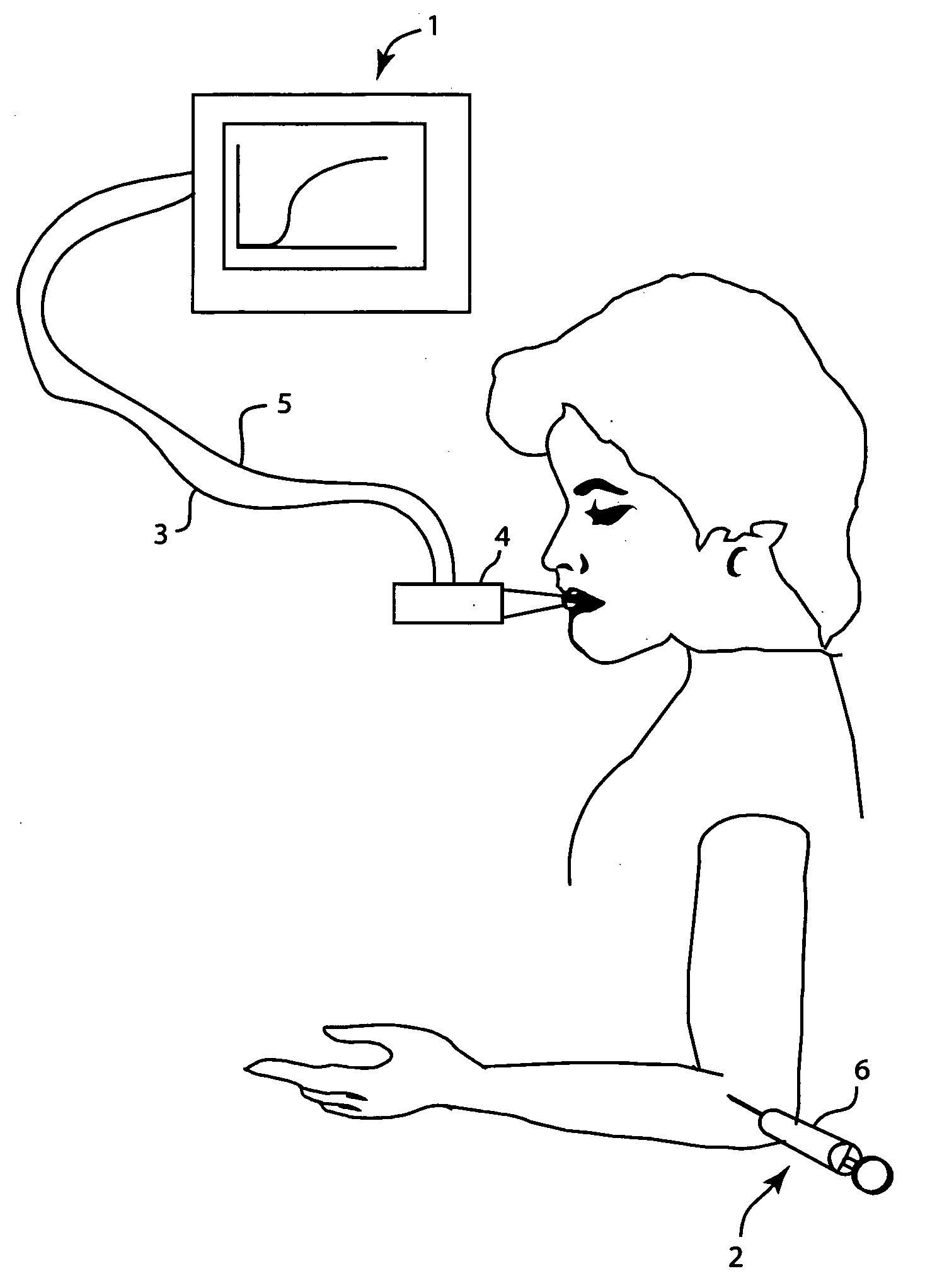
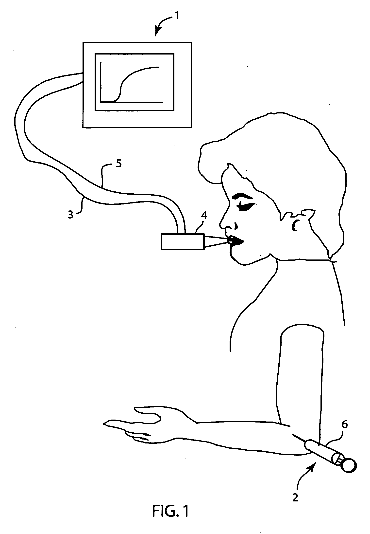
[0025]FIG. 1 shows VCap measurement apparatus 1 and arterial blood sampling apparatus 2. Apparatus 1 includes an expired breathing gas CO2 concentration sensor, which is advantageously an infrared gas analyzer. Such analyzer may be either of a mainstream type, in which the infrared absorption path is directly at the breathing gas pathway, or alternatively of a sidestream type, in which a sample of the breathing gas is withdrawn with a sampling line 3 transporting a sample flow of the breathing gas to the infrared absorption path of the sensor for measurement.

[0026] Breathing gas volume may be measured advantageously with any type of well-known flow sensor 4, based on pressure difference measurement over a known flow restrictor, a thermal sensor, an ultrasound sensor, or other suitable sensor. Flow sensor 4 is coupled to apparatus 1 by conductor 5. The volume is determined by integration of the flow signal with respect to time. If the gas concentration is determined with sidestream ...


PUM


Abstract

A method and apparatus for determining the presence or absence of a pulmonary embolism (PE) in a patient. The breathing gas CO2 partial pressure (PCO2) during the expiration of breathing gases by the patient, the end tidal (EtCO2), CO2 partial pressure, and the CO2 partial pressure (PaCO2) of the blood are measured. The volume (V) of breathing gases expired during the expiration of breathing gases by the patient is also measured and a relationship between changes in breathing gas CO2 partial pressure (PCO2) and changes in breathing gas volume (V) in an alveolar expiration phase of patient expiration is determined. The difference between the blood CO2 partial pressure (PaCO2) and the end expiration CO2 partial pressure is divided by the relationship between PCO2 and V produce a quantity which is compared to a threshold value. If the quantity is below the threshold value, the absence of a pulmonary embolism is indicated.

Description

FIELD OF THE INVENTION [0001] The present invention relates to a method and apparatus for indicating an embolic condition of a patient. To this end, the invention may be used to indicate the absence of a pulmonary embolism in a patient. BACKGROUND OF THE INVENTION [0002] Pulmonary embolism (PE) is a blockage or occlusion of a pulmonary blood vessel. Most commonly it is caused by a blood clot or thrombosis, that is, an “embolus”, in a vessel. It is a common illness with an annual incidence of 1 in a 1,000 in the Western world population. Mortality of PE is 30% when left undiagnosed and untreated but with treatment this can be reduced to 5-8%. The diagnosis of PE is difficult and is typically based on multi-step algorithms starting from an evaluation of the clinical probability and laboratory tests for markers, but positive diagnosis always requires some kind of imaging, ventilation-perfusion lung scintigraphy, pulmonary angiography, or multi-detector spiral X-ray computed tomography....

Claims


Application Information

Patent Timeline
Patent Type & Authority Applications(United States)
IPC IPC(8): A61B5/08
CPCA61B5/0836
Inventor HEINONEN, ERKKICOFFENG, RENEVERSCHUREN, FRANCK
Owner GENERAL ELECTRIC CO
Why Eureka
Try Eureka