Blood flow image diagnosing device

a technology of blood flow rate and imaging device, which is applied in the field of blood flow rate imaging device, can solve the problems of difficult to distinguish the waveform of a thin arterial blood vessel, the value has intrinsically much noise, and the statistical error occurs, so as to achieve the effect of accurately distinguishing an artery from a vein and being effective in diagnosing arteriosclerosis

Active Publication Date: 2011-12-29
NAT UNIV CORP KYUSHU INST OF TECH (JP)
View PDF17 Cites 37 Cited by
  • Summary
  • Abstract
  • Description
  • Claims
  • Application Information

AI Technical Summary

Benefits of technology

[0129]Using the device of the present invention, it becomes possible to separate a blood flow waveform of a retina vessel from one of a background tissue so as to analyze them, though it has not been possible to separate them clearly until now. Further, it is possible to distinguish an artery from a vein more accurately by grouping data along a course of each blood vessel extending circumferentially from an artery and a vein in the center. Also, in the case that a blood flow waveform has a secondary peak or in the case of a region having a highly-dispersive and complicated blood flow waveform which generally contains many points deviated greatly therefrom, it is possible to obtain a map wherein a surface layer blood flow or a retina vessel is distinguished from a background blood flow or a blood vessel of background tissue.
[0130]Conventionally, it has not been possible to measure a diameter of meandering retina vessels by setting a rubber band thereto. However, by determining a center line of blood vessel cut out using a specified mask and, subsequently, measuring a distance to an edge of the mask therefrom, it is possible to presume an inside diameter of meandering blood vessels and thereby it is also possible to evaluate a blood circulation effect. Further, since it is possible to separate an intravascular portion from a background tissue so as to evaluate each blood flow waveform and blood flow volume, there can be provided new information for evaluating functions of the circulatory system. In particular, an accurate observation of a blood flow waveform of an artery is considered extremely effective for diagnosing arteriosclerosis.

Problems solved by technology

These grains arise from facts that a speckle image for calculating a blood flow value has intrinsically much noise and a statistical error occurs since samples for measuring blood flow of each pixel are limited in number.
Therefore, it has been a big problem to develop a method for distinguishing surely a region of blood vessel running on a surface, i.e., the retina vessel region, from a region of background blood flow.
Consequently, when the distribution of skewness is displayed as an image, there is a problem that a waveform of a thin arterial blood vessel is difficult to distinguish, since it is buried in a waveform of tissue blood flow existing in the background.
As stated above, however, there has been a problem that it is difficult to find a method for extracting only blood vessels.
However, since most retina vessels are not straight as shown in FIG. 1, only limited blood vessels can be measured by the method setting up the rectangular region.
In addition, very complicated work is required to set accurately a rectangular region on a blood vessel every time blood flow analysis is performed.
Accordingly, it has been a great problem to develop a method in which a meandering blood vessel can be analyzed as well as a straight blood vessel.
Therefore, it is insufficient to characterize a blood flow waveform by only skewness, and thus it is also necessary to introduce other indices so as to judge holistically.

Method used


Image

  • Blood flow image diagnosing device
  • Blood flow image diagnosing device
  • Blood flow image diagnosing device

Examples


Embodiment Construction

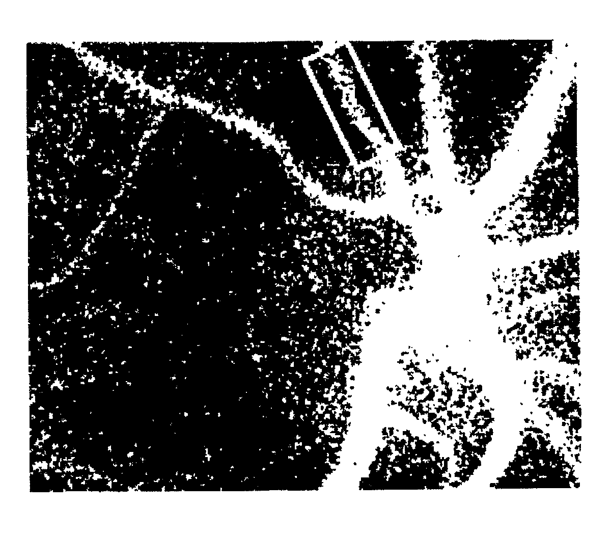
[0147]A blood flow image diagnosis device of the present invention comprises a laser beam irradiation system that irradiates a body tissue having a blood cell with a laser beam;

[0148]a light receiving system having a light receiver including a large number of pixels that detects reflected light from the body tissue;

[0149]an image capture section that continuously captures a plurality of images for a specified time that is one or more cardiac beats on the basis of a signal from the light receiver;

[0150]an image storage section that stores the plurality of images;

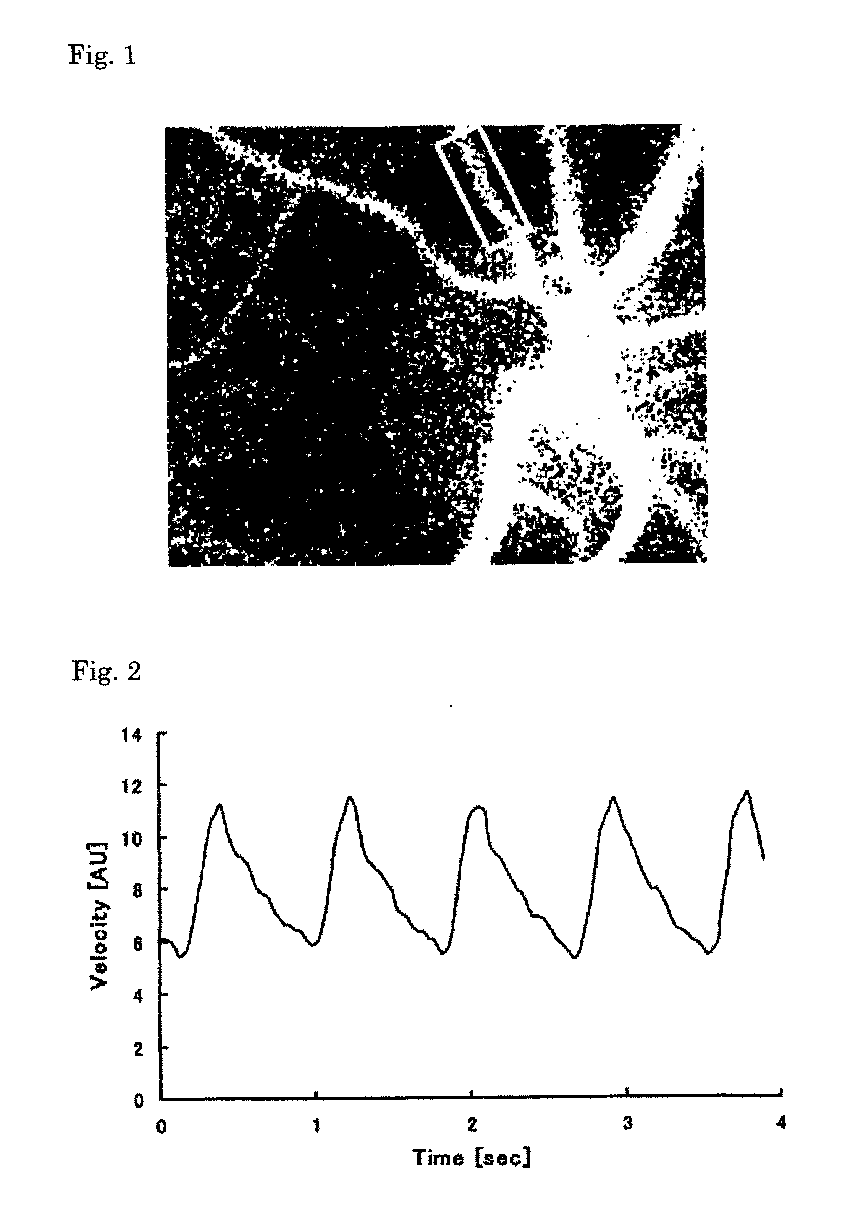
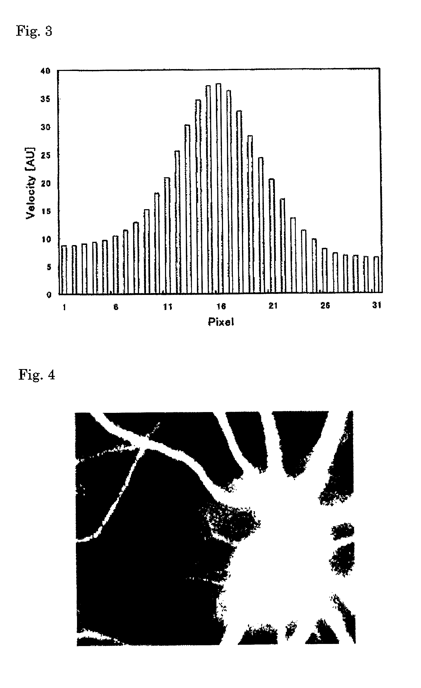
[0151]an arithmetic section that calculates a blood flow rate within the body tissue from the time variation of the output signal of each pixel corresponding to the plurality of the stored images; and

[0152]a display section that displays the two-dimensional distribution of the calculation result as a blood flow map.

[0153]Further, the arithmetic section of the blood flow image diagnosis device contains an additional function t...


PUM


Abstract

The present invention provides a blood flow image diagnosis device comprising: a laser beam irradiation system that irradiates an observation region of a body tissue with a laser beam; a light receiving system having a light receiver including a large number of pixels that detects reflected light from the observation region of the body tissue; an image capture section that continuously captures a plurality of images for a specified time that is one or more cardiac beats on the basis of a signal from the light receiver; an image storage section that stores the plurality of images; an arithmetic section that calculates a blood flow rate in the body tissue from the time variation of the output signal of each pixel corresponding to the plurality of the stored images; and a display section that displays the two-dimensional distribution of the calculation result as a blood flow map; wherein the blood flow image diagnosis device has a function for analyzing the blood flow map, the arithmetic section has a function that separates a blood flow of a blood vessel observed in a surface layer in the observation region of the body tissue from a peripheral background blood flow on the basis of a plurality of blood flow map data for a time that is one or more cardiac beats, and the display section has a function that distinguishably displays each blood flow on a blood flow map. The blood flow image diagnosis device has an additional function for diagnosing blood flow images as compared to a conventional device.

Description

TECHNICAL FIELD[0001]The present invention relates to a blood flow rate imaging device for measuring and imaging a blood flow rate on the basis of a speckle signal reflected from a body tissue obtained by irradiating the body tissue having blood cells with a laser beam wherein a new function for analyzing a blood flow imaging is added thereto.BACKGROUND ART[0002]Conventionally, the present inventors have developed a blood flow rate measuring device that irradiates with a laser beam a body tissue having blood cells such as an eye ground and a skin, leads random speckled patterns, i.e. speckle images, which are formed by interference of reflected light from the blood cells, onto an image sensor such as a solid-state image sensor (CCD or CMOS), sequentially captures and stores a large number of these speckle images at predetermined time intervals, selects a predetermined number of images from the large number of the stored images, calculates a value reflecting a time variation speed of...

Claims


Application Information

Patent Timeline
Patent Type & Authority Applications(United States)
IPC IPC(8): A61B5/02
CPCA61B3/1233A61B5/489A61B5/0261A61B5/02007
Inventor FUJII, HITOSHIOKAMOTO, KENJITAKAHASHI, NORIYOSHIISHIHARA, HIROYUKI
Owner NAT UNIV CORP KYUSHU INST OF TECH (JP)
Why Eureka
Try Eureka