
Thermally conductive surgical probe

a surgical probe and thermal conductivity technology, applied in the field of medical devices, can solve the problems of prolonged healing time, considerable discomfort, pain,

Inactive Publication Date: 2007-03-06
BALZANO ALFIERO
View PDF6 Cites 3 Cited by
  • Summary
  • Abstract
  • Description
  • Claims
  • Application Information

AI Technical Summary

Benefits of technology

[0008]Therefore, it is among the primary objects of the present invention to provide a surgically thermal conductive probe which is useful in a surgical procedure to define, identify, and provide a safe, disposable means that doctors can use to insert, position, contact and terminate, by freezing or cauterizing, an undesired internal area or body organ.
[0009]Another object of the present invention is to provide a surgical thermal conductive probe which not only will provide the above solution but also includes means for removal of frozen or cauterized infected areas, eliminating or minimizing the spread of associated infected or undesirable cells.
[0010]Still another object of the present invention is to provide a surgical procedure and thermally conductive probe which can be inserted through the smallest possible entry in the skin and which can be removed through the entry, allowing for a minimum recovery time, in comparison to present day methods.
[0012]Still a further object resides in providing a thermal composite material for a surgical probe, whereby a surgeon can activate thermal conductivity of an extreme nature to the area of infection, tumor or body organ, while restricting the movement of the probe and precluding infected cells from spreading to other body areas.

Problems solved by technology

In the past, it has been a typical practice in the medical field to remove a tumor by employing surgical methods which require extensive invasive procedures and which cause considerable discomfort, pain, and protracted healing time.

Method used


Image

  • Thermally conductive surgical probe
  • Thermally conductive surgical probe

Examples


Embodiment Construction

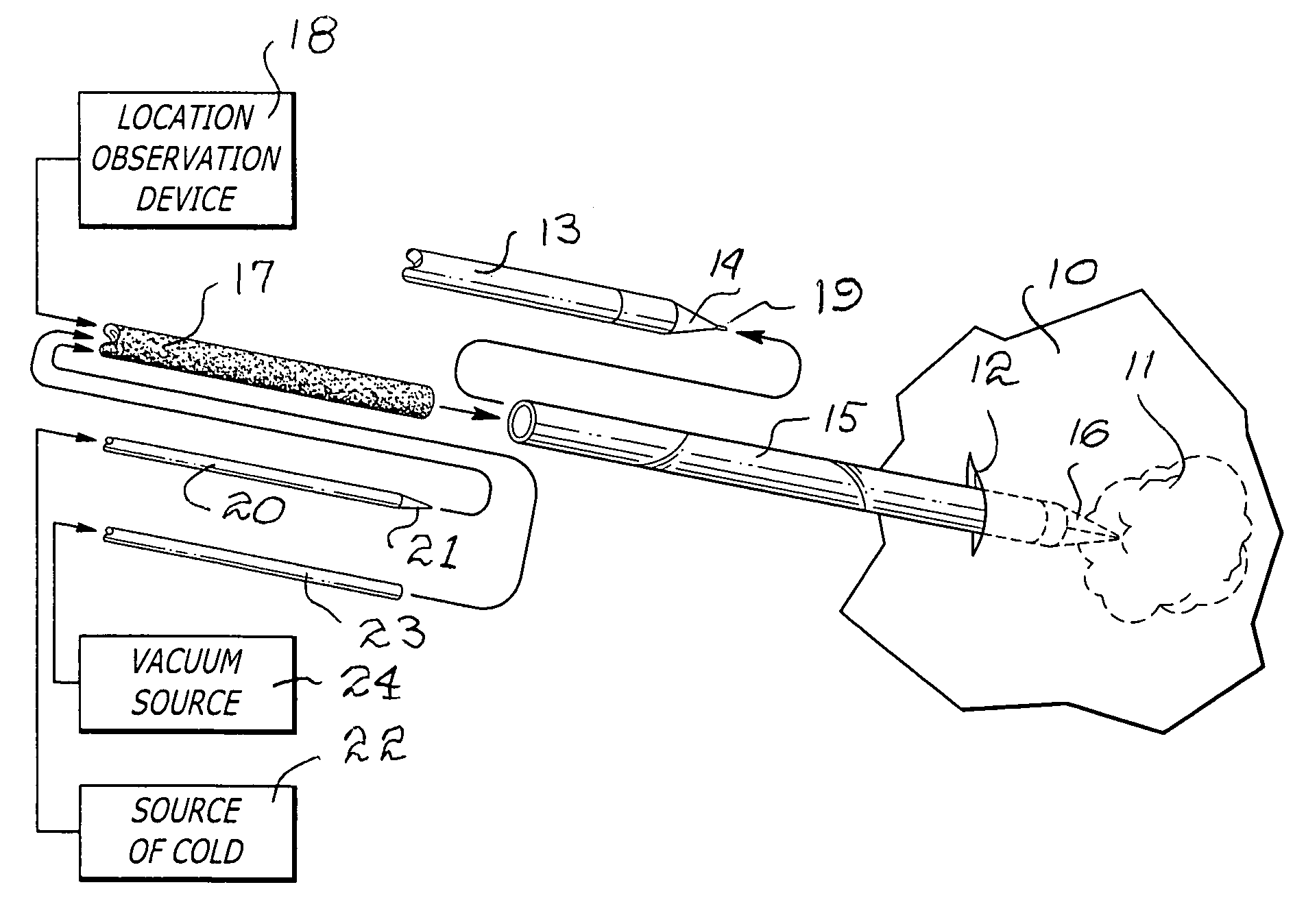
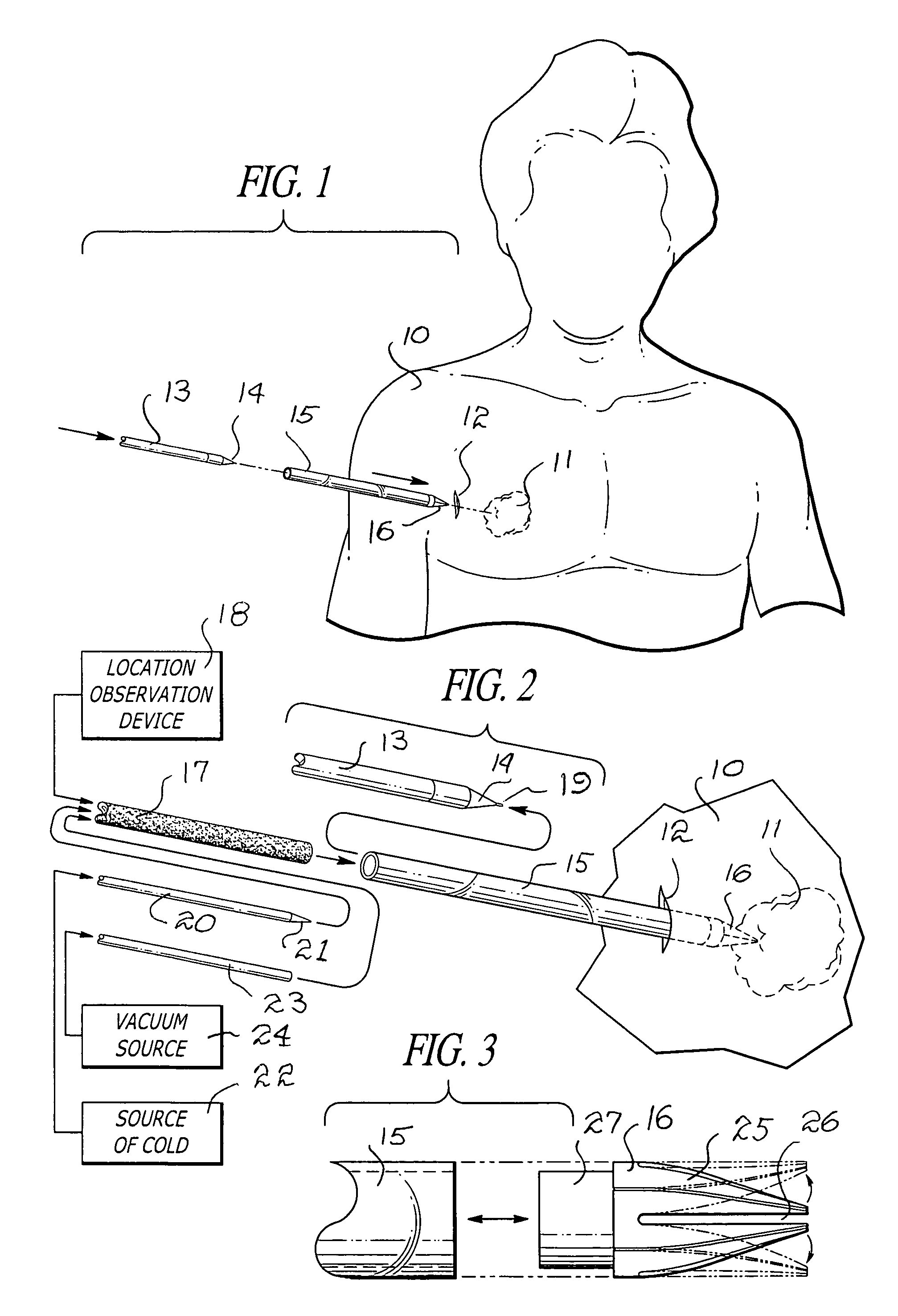
[0017]Referring now to FIG. 1, a patient is indicated by the numeral 10 having an infected area or tumor 11 which requires surgery for achieving removal. In accordance with use of the present invention, a slight incision is made in the vicinity of the area 11, and the incision is indicated by numeral 12. A steel rod 13 having a pointed or conical end 14 is inserted through the open end of a sleeve 15 and advanced through the sleeve 15 so that the conical end 14 terminates within a flapped conical end 16 of the sleeve. Subsequently, the sleeve and the rod 13 are inserted simultaneously through the incision and advanced through surrounding tissue in the direction of the infected area or tumor 11.

[0018]Referring to FIG. 2, it can be seen that the rod within sleeve 15 is advanced until an extension 19 at the tip of the rod is immediately adjacent to the infected area or tumor 11. At this time when the area to be worked upon is located, the rod 13 is removed from the sleeve 15 and replac...


PUM


Abstract

A surgical apparatus and process for freezing or cauterizing a particular infected body area employing a transparent sleeve insertable through a small slit in the flesh of a patient and wherein the inserted end of the sleeve is provided with a cone-shaped tip having a plurality of segments normally self biased to close to define a dull pointed end susceptible for expansion when a solid, rigid probe is inserted through the sleeve. The end of the probe includes a conical or cone-shaped tip with a small extension or projection that causes expansion of the flaps on the tip of the sleeve. The extension on the end of the probe is placed in alignment position to locate the infected organ or area to be removed. Monitoring of the sleeve and probe insertion and location of the infected area is achieved by video camera. After removing the solid and rigid probe, a fiber optic tube is inserted into the sleeve whereby ambient light is conducted through the tube to the advancing end of the sleeve. A carbon rod is inserted through the fiber optic tube so as to have its pointed end in contact with the tumor, body organ or infected area. The carbon rod is connected to a source of cold or heat. In a cold application, the infected area or tumor is frozen so that when the carbon rod is removed and replaced by a suction device, the frozen infected area or tumor can be fragmented and the particles removed.

Description

[0001]Priority claimed based on Ser. No. 60 / 440,098 filed Jan. 16, 2003 pending.BACKGROUND OF THE INVENTION[0002]1. Field of the Invention[0003]The present invention relates to the field of medical devices and more particularly to a novel surgical probe and process for medical use, whereby a thermal conductive probe is introduced to a diseased or tumor affected area of the body, whereby the infection or tumor is solidified sufficiently so that subsequent removal of hardened particles, cells or the like can be withdrawn by suction means.[0004]2. Brief Description of the Prior Art[0005]In the past, it has been a typical practice in the medical field to remove a tumor by employing surgical methods which require extensive invasive procedures and which cause considerable discomfort, pain, and protracted healing time. Therefore, a long-standing need has existed to provide a surgical device and method which would readily define, identify, and provide a safe, disposable probe solution where...

Claims


Application Information

Patent Timeline
IPC IPC(8): A61B18/18
CPCA61B18/02A61B2218/008A61B18/04A61B2017/3454A61B90/40
Inventor BALZANO, ALFIERO
Owner BALZANO ALFIERO
Learn More