SPECT examination device

a spectrometer and spectrometer technology, applied in the field of single photon emission computed tomography, can solve the problems of inability to compute single photon emission tomography, the sensitivity of the device declines in a disadvantageous manner, etc., and achieves the effects of enhancing the positional resolution, and improving the positional resolution

Inactive Publication Date: 2007-04-03
SCIVIS +1
View PDF7 Cites 16 Cited by
  • Summary
  • Abstract
  • Description
  • Claims
  • Application Information

AI Technical Summary

Benefits of technology

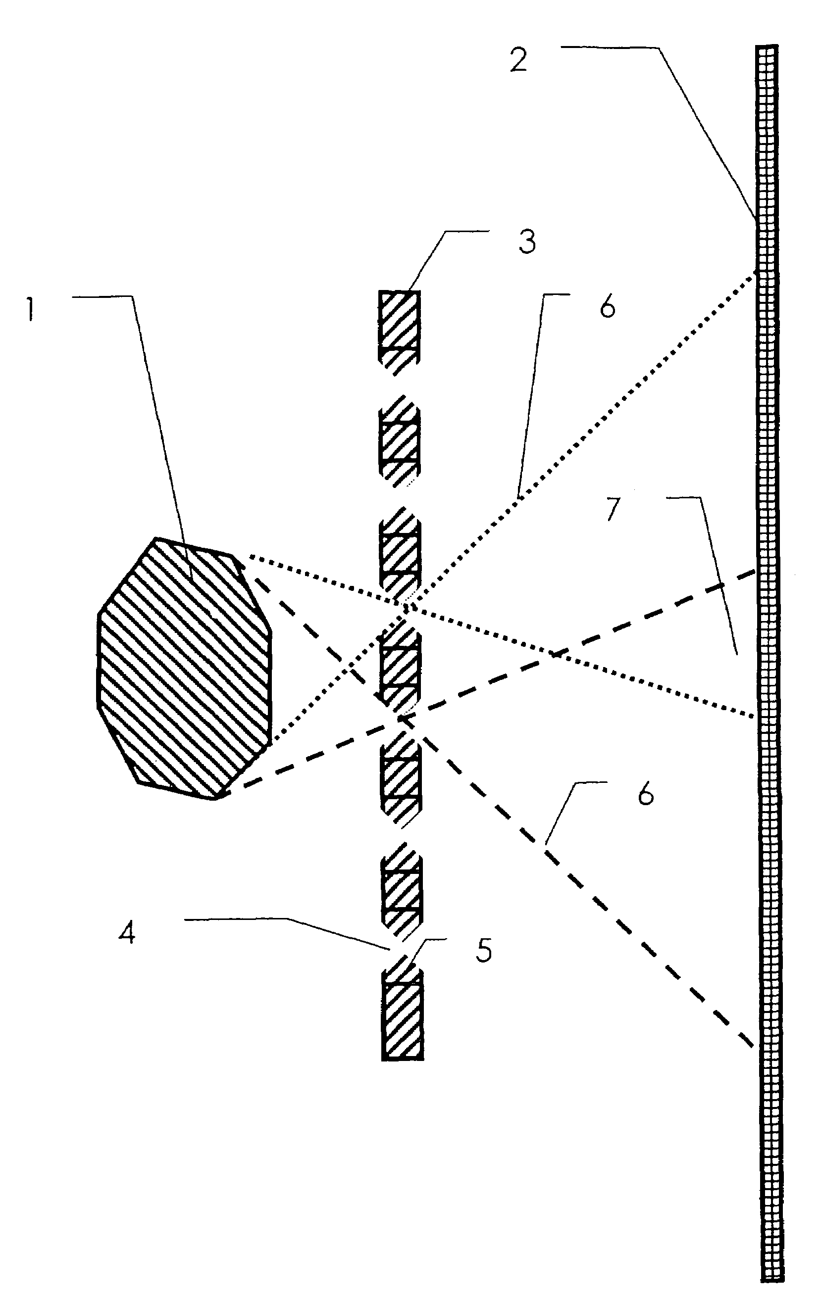
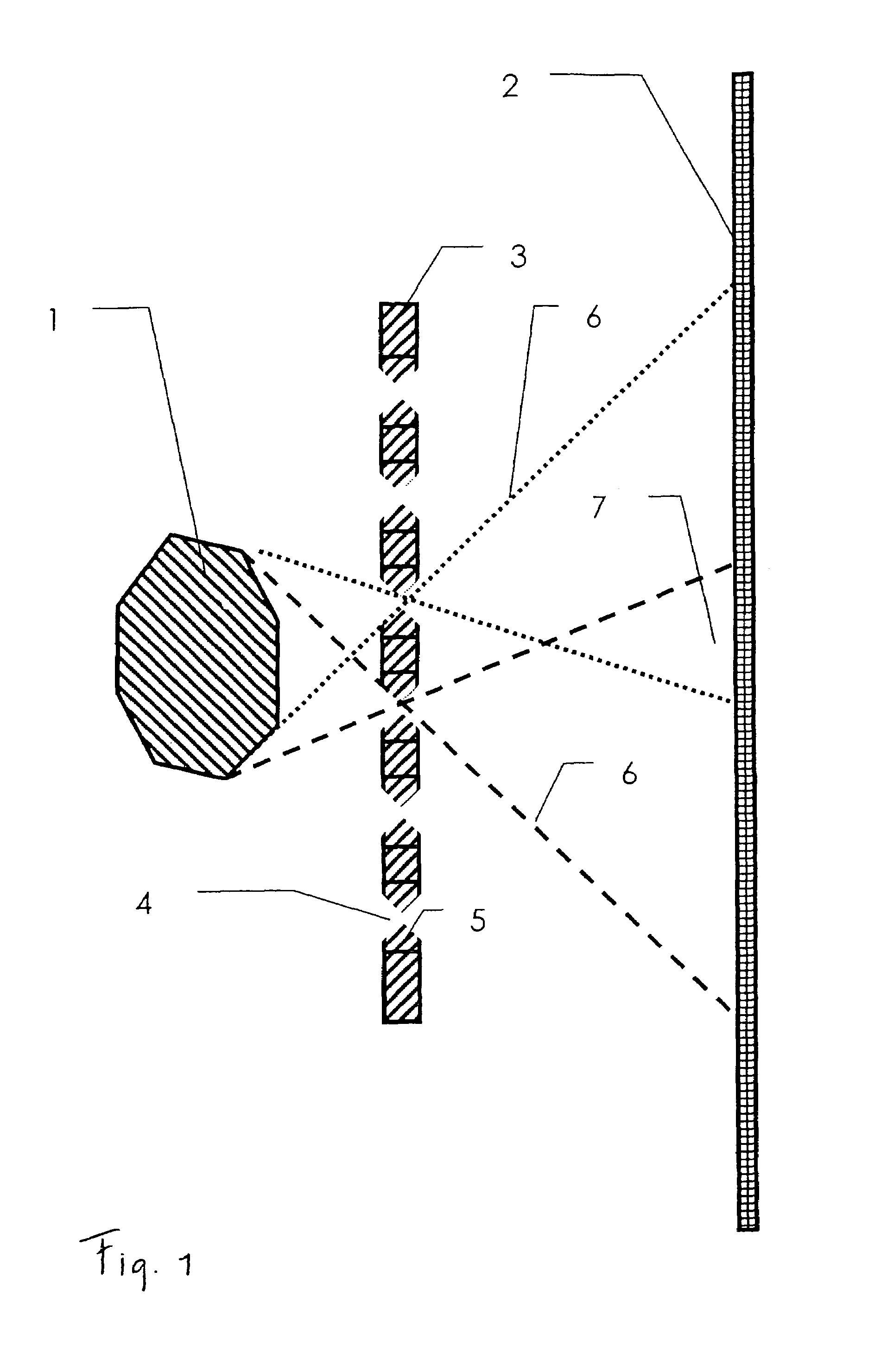
[0009]To improve the positional resolution by comparison with the prior art named above, a pinhole collimator is used in single photon emission computed tomography. A pinhole collimator is characterised by a single pinhole, through which the photons pass. If the object is closer to the pinhole collimator than the surface of a gamma camera and / or of a detector, this will ultimately achieve an enlarged positional resolution. The photons do not pass through the pinhole collimator exclusively in an perpendicular direction. Instead, they enter and leave in a conical shape. Since the cone located behind the pinhole collimator is larger than the cone in front of the pinhole collimator, an improvement in positional resolution is achieved by comparison with the prior art named in the introduction.

Problems solved by technology

Accordingly, as the pinholes become smaller, the sensitivity of the device declines in a disadvantageous manner.
If the sensitivity is too low, single photon emission computed tomography is ultimately no longer possible.

Method used


Image

  • SPECT examination device
  • SPECT examination device
  • SPECT examination device

Examples


Embodiment Construction

[0013]The object of the invention is to create a device with an associated method of the type named above, which allows high-resolution and high-sensitivity measurements.

[0014]The device claimed comprises a multiple-pinhole collimator together with a detector for registering photons which pass through the multiple-pinhole collimator. Accordingly, the collimator provides a large number of pinholes. In one embodiment of the invention, the detector is designed in such a manner that it can also measure the energy of the photons detected.

[0015]Since the collimator provides several pinholes, the sensitivity of the device is increased accordingly. The use of a pinhole collimator, by comparison with the use of collimators which can only register perpendicular beams falling in a perpendicular direction, provides the advantage of a high positional resolution. Accordingly, a device with good positional resolution and good sensitivity is provided.

[0016]During the operation of the device, the ob...


PUM


Abstract

A tomography device, particularly for single photon emission computed tomography (SPECT), comprises a multi-pinhole collimator and a detector for detecting gamma quanta or photons that penetrate the multi-pinhole collimator. In a tomographic method using the device, the distance between the object and the multi-pinhole collimator is selected to be smaller than the distance between the multi-pinhole collimator and the surface of the detector.

Description

SPECT EXAMINATION DEVICEBACKGROUND OF THE INVENTION AND RELATED ART[0001]The invention discloses a device and a method for tomography, especially single photon emission computed tomography (SPECT).[0002]Single photon emission computed tomography is a method with associated devices for three-dimensional imaging of radio pharmaceuticals, which have been introduced into an object. Human beings or animals may be provided as the object. The radio pharmaceuticals introduced into the object emit photons or gamma quanta. These photons are registered and evaluated by the device. The position, that is to say, the spatial distribution of the radio pharmaceuticals, is obtained as the result of this evaluation. In turn, the position of the radio pharmaceuticals allows inferences to be made regarding the object, for example, regarding a distribution of tissue in the object.[0003]One known device for the implementation of single photon emission computed tomography comprises a gamma camera and a co...

Claims


Application Information

Patent Timeline
Patent Type & Authority Patents(United States)
IPC IPC(8): G01T1/164
CPCG01T1/1648
Inventor SCHRAMM, NILSHALLING, HORSTEBEL, GERNOT
Owner SCIVIS
Why Eureka
Try Eureka