Color space transformations for use in identifying objects of interest in biological specimens

a color space and object technology, applied in image enhancement, image data processing, instruments, etc., can solve the problems of ratio-based color transformation, difficult detection of cancer cells or particular proteins in biological specimens, improper characterization of areas, etc., to achieve the effect of observing and identifying objects

Inactive Publication Date: 2007-04-03
VENTANA MEDICAL SYST INC
View PDF24 Cites 31 Cited by
  • Summary
  • Abstract
  • Description
  • Claims
  • Application Information

AI Technical Summary

Benefits of technology

[0013]Methods are provided herein for identifying objects of interest, which take advantage of using one or more novel color space transformations described herein. Several methods are described herein for conceptualizing the color space transformations, and for carrying out the transformations in practice using a general-purpose computer. The methods described below are preferably coded in software as a set of instructions for the general-purpose computer. The methods are particularly designed for use in processing an image having red green and blue pixel components and transforming that image to another representation with the computer and displaying the transformed image to the user. The transformed image enables a human operator (e.g., pathologist or technician) to more readily observe and identify objects of interest that are contained in specimen.
[0014]The first transformation, referred to herein as “Minus Clear Plus One” or “MC+1”, is suited to staining methods that produce at least two different colors. Conceptually, the MC+1 transformation involves translating and rotating axes of a three-dimensional coordinate space that defines an image of the biological specimen. Alternatively, the MC+1 transformation involves calculating differences between vectors in the three dimensional coordinate space. The MC+1 transformation has a high degree of sensitivity to objects of interest, while being insensitive to characterizing areas of intense staining or foreign debris produced as a result of the staining as objects of interest. Therefore, the MC+1 transformation does not produce false identification of objects of interest, typical of the ratio-based color transformation.
[0015]The other of the color space transformations is referred to herein as the “Quantitative Chromatic Transformation” or “the QCT”. Like the MC+1 transformation, the QCT is also sensitive to identifying objects of interest, but insensitive to characterizing areas of intense staining or foreign debris produced as a result of the staining as objects of interest. The QCT quantitates, for each pixel, a number of absorbing molecules. The QCT produces quantities that are linearly related to concentration of analytes and is robust in the presence of instrument calibration errors. The QCT has a high specificity to colors of interest in the biological specimen and does not introduce quantitation errors, also typical of the ratio-based color transformation.
[0016]Application of one, or both, of the above two color transformations to the image of the biological specimen results in a transformed image (or transformed images if both are used) that aid in the identification of the objects of interest in the biological specimen.
[0021]The Quantitative Chromatic Transform is a colorimetric transformation that produces three new quantitities from the original red, green, and blue pixel values for each color pixel of an image. These three new quantities, X, Y, and Z can each be easily related to the quantitative amount of absorbing molecules sampled by that pixel by the logarithm function. The distinctive feature of the QCT, is its ability to quantitate analytes from color image data.

Problems solved by technology

The cancer cells or the particular proteins in the biological specimen can be difficult to detect.
The areas of intense staining and foreign debris can cause the ratio-based color transformation of the '892 patent to improperly characterize the areas of intense staining or foreign debris as objects of interest.
Since the components of the pixels in such areas are relatively “low,” and since low components that make up such pixels can appear in the denominator of the ratios, the color ratio can have falsely high values, causing “false” objects of interest to appear.
Like the MC+1 transformation, the QCT is also sensitive to identifying objects of interest, but insensitive to characterizing areas of intense staining or foreign debris produced as a result of the staining as objects of interest.

Method used


Image

  • Color space transformations for use in identifying objects of interest in biological specimens
  • Color space transformations for use in identifying objects of interest in biological specimens
  • Color space transformations for use in identifying objects of interest in biological specimens

Examples


Embodiment Construction

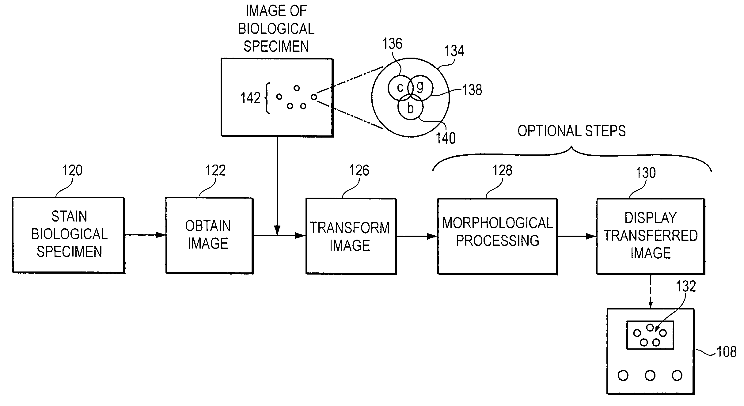
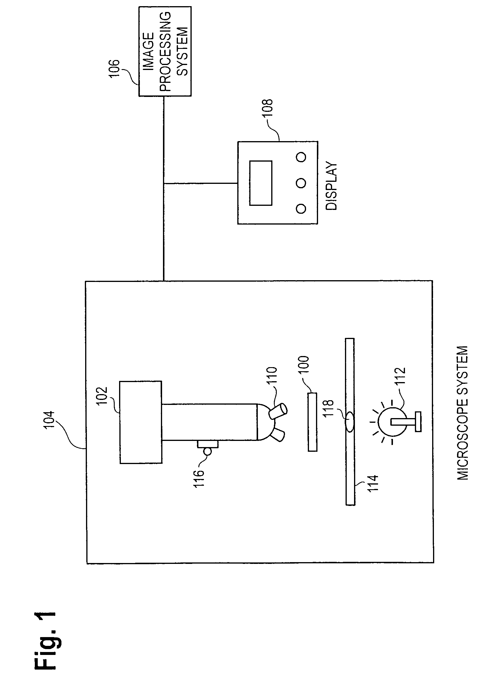
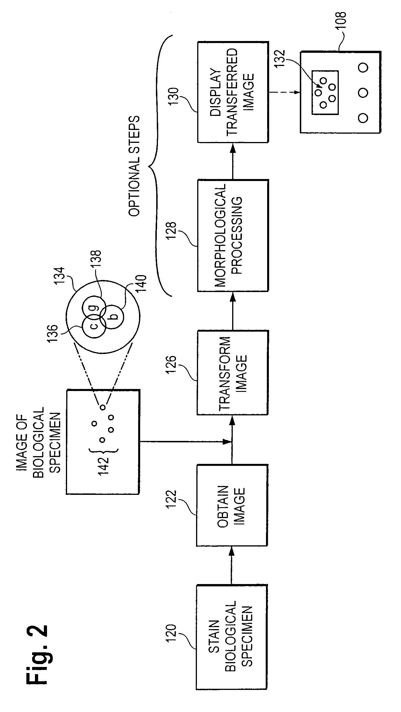
[0036]FIG. 1 is a block diagram of exemplary apparatus for automated cell analysis of a biological specimen, in which exemplary embodiments of the present invention may be employed. It should be understood that this and other arrangements and elements (e.g., machines, interfaces, functions, orders of elements, etc.) can be added or used instead and some elements may be omitted altogether. Additionally, those skilled in the art will appreciate that many of the elements described herein are functional entities that may be implemented as discrete components or in conjunction with other components, in any suitable combination and location. Moreover, the various functions described herein as being performed by one or more entities may be carried out by hardware or by a processor programmed to execute an appropriate set of computer instructions stored in memory. Provided with the present disclosure, those skilled in the art can construct the hardware and develop the appropriate set of com...


PUM

PropertyMeasurementUnit
color space transformationsaaaaaaaaaa
coloraaaaaaaaaa
colorsaaaaaaaaaa

Abstract

Two color transformations, as described herein, facilitate identification of the objects of interest in the biological specimen. One of the color transformations, a Minus Clear Plus One (MC+1) transformation, can be conceptualized as either translating and rotating axes of a three-dimensional coordinate space that defines an image of the biological specimen or calculating differences between vectors in the three dimensional coordinate space that defines the image of the biological specimen. The other of the color transformations, a Quantitative Chromatic Transformation (QCT), is a colorimetric transformation that produces three new quantitities from the original red, green, and blue pixel values for each color pixel of an image. These three new quantities, X, Y, and Z can each be related to the quantitative amount of absorbing molecules sampled by that pixel. Application of one or both of the color transformations to the image of the biological specimen results in a transformed image, in which objects of interest are more readily identifiable.

Description

BACKGROUND[0001]1. Field of the Invention[0002]This invention relates generally to the field of identification and analysis of biological specimens, and more particularly to a method and system for identifying objects of interest, such as cancerous cells or cellular objects, in a biological specimen. The invention is also related to the field of color space transformations, in which a representation of an object in one color space (e.g., red, green and blue components) is transformed mathematically into a new representation in a new color space, in order to more easily observe or identify objects.[0003]2. Description of Related Art[0004]A biological specimen such as samples of bone marrow, cervical tissue, lymph nodes, or peripheral blood, may have objects of interest to a pathologist or histologist. Such samples are typically fixed to a slide and examined under a microscope. An important aspect of medical diagnostics is detecting, identifying, and quantitating the objects of intere...

Claims


Application Information

Patent Timeline
Patent Type & Authority Patents(United States)
IPC IPC(8): G06K9/00G01N33/48G01N15/14G01N33/483G06T7/00
CPCG01N15/1475G06T7/0012G06K9/00127G01N2015/1472G06T2207/30004G06V20/69
Inventor DOUGLASS, JAMES
Owner VENTANA MEDICAL SYST INC
Why Eureka
Try Eureka