Image sensor for dental intraoral radiography

Inactive Publication Date: 2007-10-16
GENDEX +1
View PDF13 Cites 33 Cited by
  • Summary
  • Abstract
  • Description
  • Claims
  • Application Information

AI Technical Summary

Benefits of technology

[0009]The present invention has the object of solving the above problems at least in part. In particular, the intraoral sensor of the present invention should enable good patient cooperation, thus making it possible to take high qua

Problems solved by technology

The design of a dental image

Method used


Image

  • Image sensor for dental intraoral radiography
  • Image sensor for dental intraoral radiography
  • Image sensor for dental intraoral radiography

Examples


Embodiment Construction

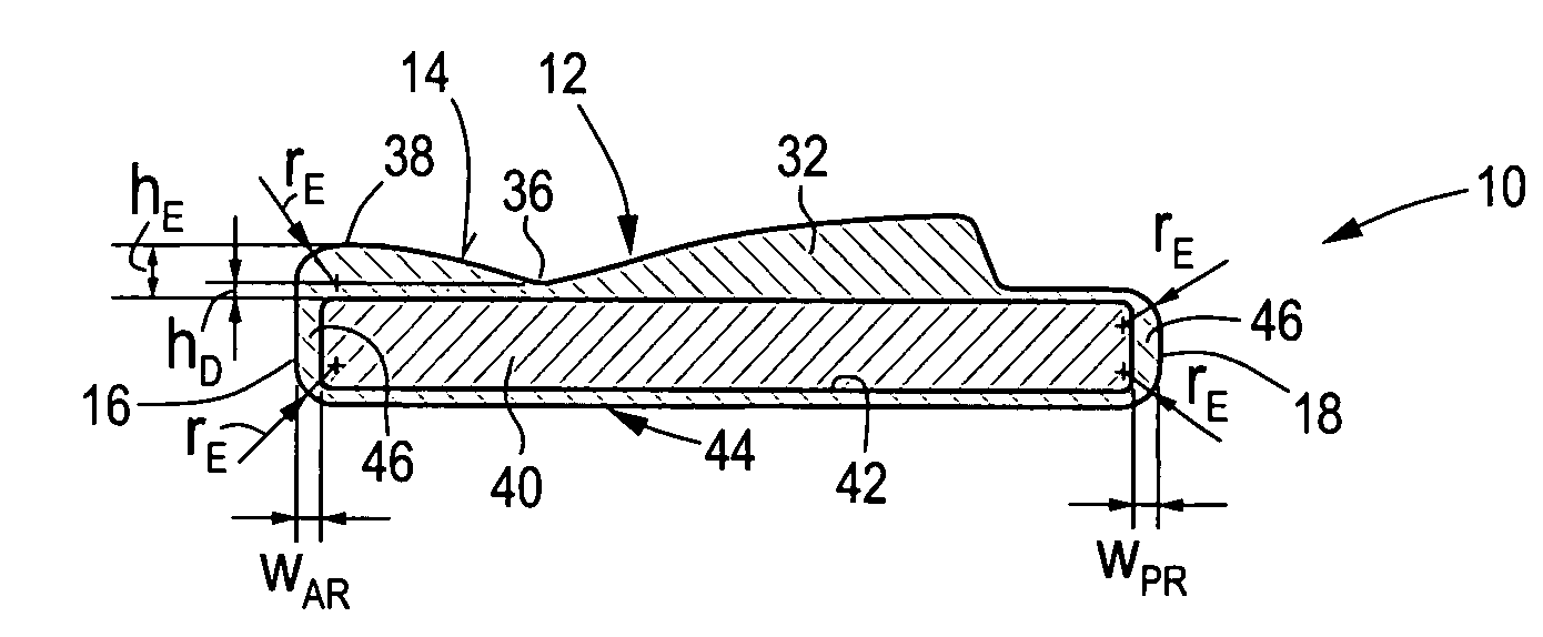
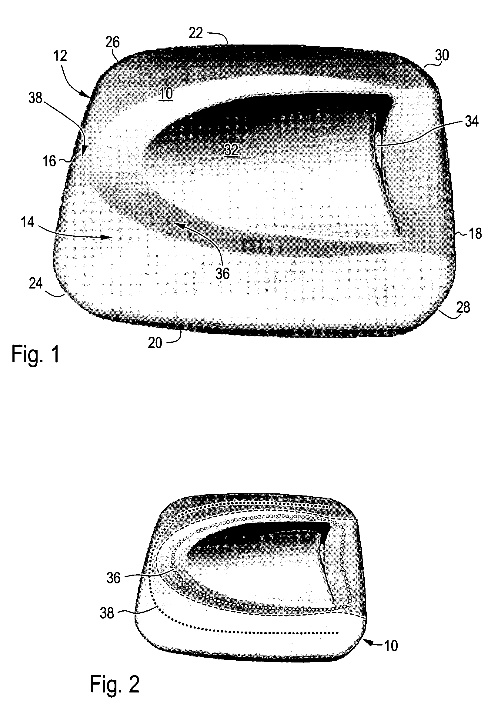
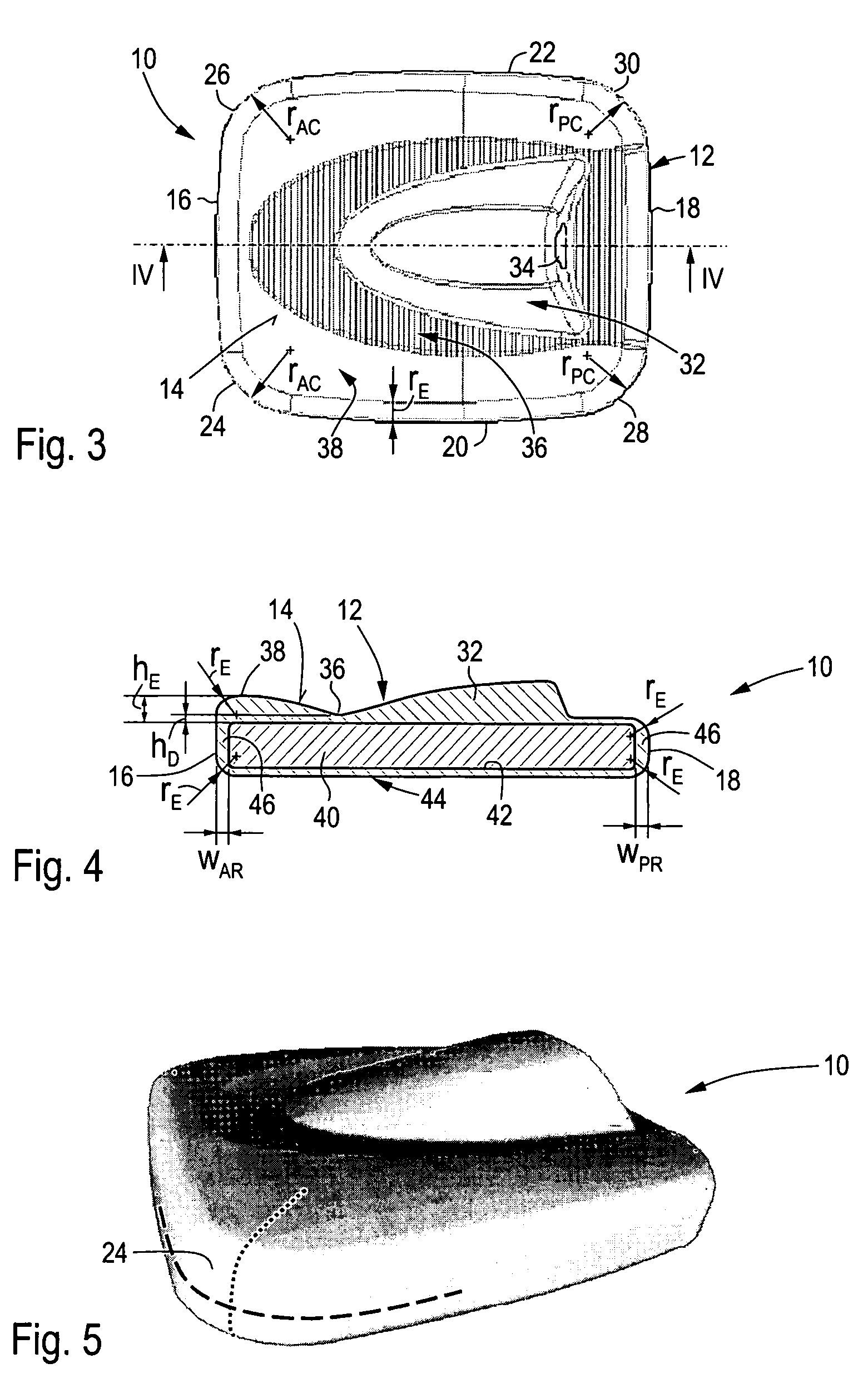
[0024]The dental image sensor 10 shown in FIG. 1 comprises a housing 12 that encapsulates an image receptor (not shown). FIG. 1 depicts a back side 14 of the housing 12, i.e., the side that is opposite to a front side (not shown). The housing 12 has an anterior side 16, a posterior side 18 and two lateral sides 20, 22. The anterior side 16 merges into the lateral sides 20, 22 at two anterior corners 24, 26. Likewise, the posterior side 18 merges into the lateral sides 20, 22 at two posterior corners 28, 30.

[0025]The back side 14 of the housing 12 has an approximately central, elongated, spade-shaped cable connection dome 32. The cable connection dome 32 comprises a cable outlet 34 that is arranged on a steep wall of the cable connection dome 32 facing towards the posterior side 18. A cable (not shown) exits the housing 12 at the cable outlet 34. The cable serves for supplying the image sensor 10 with electricity and for transmitting image information obtained by the image sensor 10 ...


PUM


Abstract

An image sensor (10) for dental intraoral radiography comprises a housing (12), an image receptor (40) and a cable connection dome (32) arranged on a back side (14) of the housing (12). The back side (14) of the housing (12) has a depression (36) at least in the region between the cable connection dome (32) and an anterior side (16) of the housing (12), wherein the depression (36) falls short of reaching the anterior side (16) of the housing (12) such that an elevated region (38) is formed between the depression (36) and at least the anterior side (16) of the housing (12). A positioning device (50) is adapted for positioning an image sensor (10) according to the present invention in the mouth of a patient. The present invention provides an intraoral sensor that enables good patient cooperation, thus making it possible to take high quality dental x-ray images.

Description

BACKGROUND OF THE INVENTION[0001]The present invention concerns the field of intraoral sensors for taking x-ray images.[0002]The first electronic intraoral x-ray image sensors have been commercialized approximately in 1990. These sensors are intended to be placed in the mouth of a patient to produce an x-ray image of the teeth and / or the jaw. The sensor typically comprises an electronic image receptor having an active imaging area. The receptor is encapsulated and protected by a housing, which may be composed of plastic and / or metal elements. Image sensors of this type of construction have rapidly gained market share over the traditional method of using photographic film because the electronic x-ray image is immediately available and can be enhanced on a computer using sophisticated imaging software.[0003]Typical dental applications of such image sensors are the acquisition of so-called “periapical” and “bite-wings” images. In order to take a periapical image, the sensor is position...

Claims


Application Information

Patent Timeline
IPC IPC(8): H01J31/49
CPCA61B6/145H04N5/32H04N5/2252H04N23/51
Inventor KOKKALIARIS, STELIOSLANGELLA, LUCIANO
Owner GENDEX
Why Eureka
Try Eureka