
Virtual telemicroscope

a virtual telemicroscope and microscope technology, applied in the field of virtual telemicroscopes, can solve the problems of pathologists hesitant to make pathologic diagnoses, microscopic images are handled and viewed in a very different way from the traditional way, and achieve the effect of different magnification levels

Inactive Publication Date: 2007-11-06
THE RES FOUND OF STATE UNIV OF NEW YORK
View PDF17 Cites 83 Cited by
  • Summary
  • Abstract
  • Description
  • Claims
  • Application Information

AI Technical Summary

Benefits of technology

[0010]In accordance with yet another exemplary aspect of the invention, a plurality of images of a specimen are captured. The images correspond to an area of the specimen and one or more segments of the specimen. The images corresponding to the specimen segments have different magnification levels; a linking map is generated between said segment images. The lin

Problems solved by technology

However, telepathology and telemedicine face a number of obstacles.
In addition, for telepathology in particular, much of the resistance to these new technologies from the pathology community has been that the microscopic images have been handled and viewed in a way that is very different from the traditional manner in which the cases are evaluated.
The pathologists frequently hesitate in making any pathologic diagnosis based on the computer images of a partial tissue section displayed in an unfamiliar manner.
When the entire specimen is digitized, the process takes a considerable amount of time (a few hours), involving specially designed and expensive automatic microscopic equipment and lengthy scanning steps.
This, together with the limited computer capacity, the relatively narrow transmission bandwidth and the very high cost, has hindered the practical value of this potentially very useful technology up to the present day.
However, no practical system was available to provide a pathologist with a fill magnified or non-magnified view of the entire specimen and at the same time a desired view of selected portions of the specimen at a different magnification.
The available systems rely on remotely controlled microscope and camera and broadband network connection, or lengthy process of digitization of slides, involving cost far beyond the justification of the practical value for most pathologists worldwide.

Method used


Image

  • Virtual telemicroscope
  • Virtual telemicroscope
  • Virtual telemicroscope

Examples


Embodiment Construction

1. Basic Principles

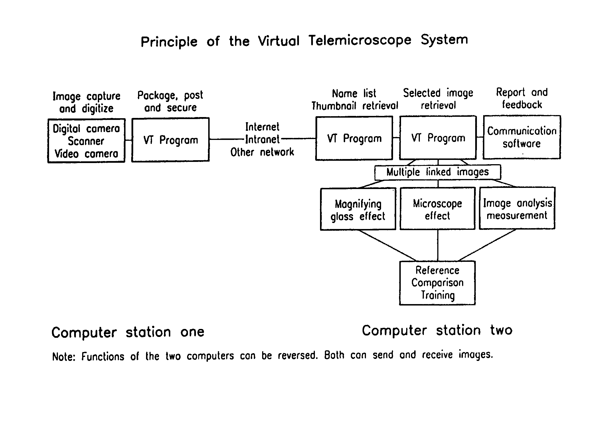
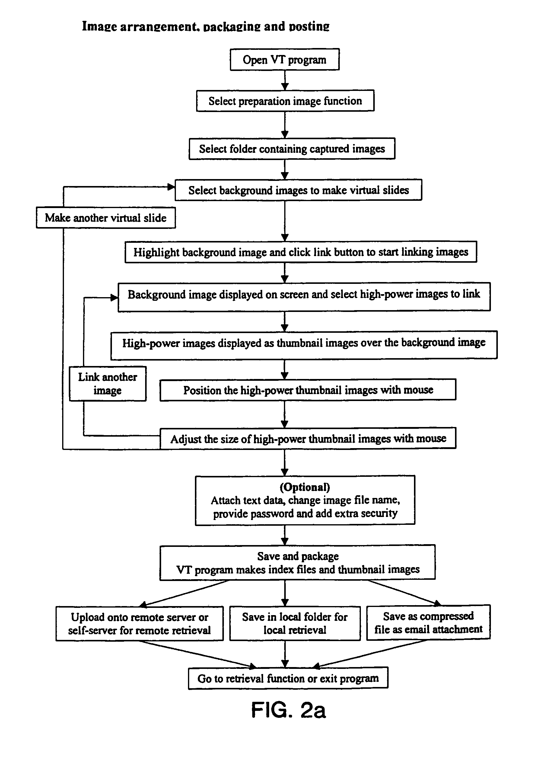
[0024]The present invention is a new “Virtual Telemicroscope (VT) system”, in which images are captured, digitized, arranged, packaged, posted, transmitted, displayed, enlarged, measured and analyzed with a user-friendly software program. It can be used for telepathology, telemedicine, distance learning, remote training, standardized exam and other applications, in which high-resolution images are transmitted and evaluated. This invention enables the users to retrieve and view virtual slides with specimen images and logically linked high-resolution partial images anywhere, any time via the Internet and other computer networks, without involving special and expensive equipment and setup.

[0025]FIG. 1 shows a functional structure of the VT system according to the present invention, which can be used to create, retrieve and view virtual slides. The basic principle of this system closely mimics that of a light microscope. When a pathologist exams a specimen on a glass ...


PUM


Abstract

A method and system that uses a computer system as a telemicroscope. A plurality of images of a specimen are captured (Digital camera scanner video camera). The images correspond to the entire specimen and a plurality of segments of the specimen (Multiple linked images). The high-resolution images corresponding to said plurality of segments have different magnification levels and locations (magnifying glass effect); a linking map is generated between said images (Multiple linked images). The linking map comprises information regarding geographical location of the images in relation to the specimen's structure (Image geographical measurement); and images and said linking map are transmitted to a remote user via a computer network thereby allowing the user to view the images with different magnification levels without compromising in image clarity (Computer station one and computer station two). The transmitted images are viewed in a dynamic manner, permitting the user to navigate, enlarge, measure, compare, annotate and exam the digitized images on a virtual slides displayed on a computer screen (Image analysis measurement). The operation of the system closely mimics that of a light microscope.

Description

BACKGROUND OF THE INVENTION[0001]This invention is related to a “Virtual Telemicroscope” (VT) system and, more particularly, to a method and system for using a computer system as a telemicroscope.[0002]Telepathology is a field that combines the disciplines of pathology, computer science and telecommunication. It captures, digitizes, transmits, displays and manipulates pathological and medical images for the purpose of analysis, consultation, collaboration, diagnosis; training and standardization. Compared with conventional pathology, telepathology is more efficient, economical and flexible. It enables medical image evaluation to be performed at any location, any time, as long as the evaluator has access to a computer with adequate network connection.[0003]The history of telemedicine and telepathology goes back to the early days of computer science and telecommunication, and its progress has paralleled the advancement of these two fields. Physicians have long been experimenting with ...

Claims


Application Information

Patent Timeline
IPC IPC(8): G06T9/00G06K9/00G06K9/36
CPCG02B21/367H04N19/29G06T9/00
Inventor GU, JIANGANDERSON, VIRGINIA M.
Owner THE RES FOUND OF STATE UNIV OF NEW YORK
Learn More