Device-less gating of physiological movement for improved image detection

a technology of physiological movement and image detection, applied in tomography, instruments, applications, etc., can solve the problems of long setup and calibration time of devices and systems for monitoring physiological movement, affecting diagnosis, and introducing errors

Active Publication Date: 2009-08-11
GENERAL ELECTRIC CO
View PDF4 Cites 26 Cited by
  • Summary
  • Abstract
  • Description
  • Claims
  • Application Information

AI Technical Summary

Problems solved by technology

Such image quality may affect the diagnosis.
Setup and calibration of the devices and systems for monitoring physiological movement can be long and complicated, and is not typically used for all exams.
Furthermore, when gating on different systems and / or at different times, errors may be introduced when attempting to plan therapy dynamically.
However, without the detection of and correction for physiological motion, incorrect diagnoses may result.

Method used


Image

  • Device-less gating of physiological movement for improved image detection
  • Device-less gating of physiological movement for improved image detection
  • Device-less gating of physiological movement for improved image detection

Examples


Embodiment Construction

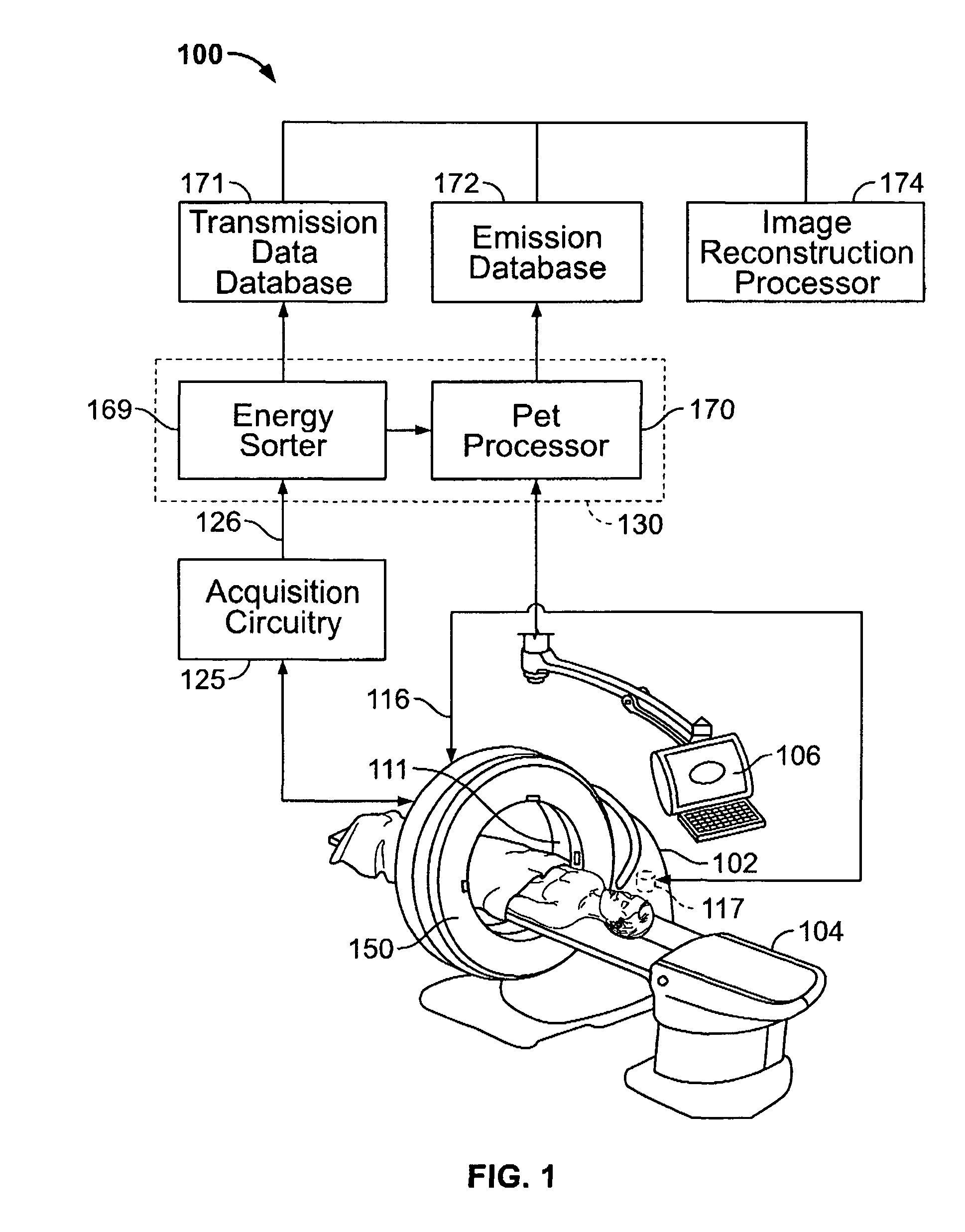
[0019]FIG. 1 is a block diagram of a medical imaging system 100 formed in accordance with an exemplary embodiment of the present invention. The system formed in accordance with an embodiment of the present invention may be any emission-type computed tomography imaging system including, but not limited to a single PET scanner, a dual PET / CT scanner, a single nuclear (photon emission) computed tomography (SPECT) scanner or a dual SPECT / CT scanner. A medical imaging system 100 such as, for example, a positron emission tomography (PET) system, includes a gantry 102, a patient table 104 and a computer system 106. Gantry 102 provides mechanical support for mounting devices such as, for example, detectors, scanners and transmitters that are useful for scanning a patient. Gantry 102 houses imaging devices such as, for example, PET detectors. The PET system is a stationary annular detector with potential variant of a pin source for PET.

[0020]In accordance with an embodiment of the invention,...


PUM


Abstract

A method of imaging an object using a medical imaging system is provided. The imaging method acquires a stream of sinogram data. Changes in the stream of sinogram data corresponding to movement of the object are detected. The method selects portions of the stream of sinogram data. An image of the object based on the selected portions of the sinogram data is generated.

Description

BACKGROUND OF THE INVENTION[0001]The present invention relates to diagnostic imaging methods and systems. In particular, the present invention relates to methods and systems for device-less gating of a stream of sinogram data.[0002]At least some known Positron Emission Tomography (PET) and Computed Tomography (CT) systems experience image quality that is highly affected by physiological patient motion. Such image quality may affect the diagnosis. Lung nodules, cardiac wall features or other small features of interest that move due to physiological motion such as cardiac and respiratory motion, may appear unfocused or faint without proper corrections. By employing a cardiac and / or respiratory gating protocol during scan acquisition, images may be classified according to physiologic position in the cardiac / respiratory cycle. The gating technique may correct for motion artifacts in images. Also, the image pathway of the nodule or other features of interest may be tracked.[0003]Respirat...

Claims


Application Information

Patent Timeline
Patent Type & Authority Patents(United States)
IPC IPC(8): A61B5/05
CPCA61B5/113A61B6/5235A61B6/037A61B6/032
Inventor PIACSEK, KELLY LYNNCHU, JANICEKOHLMYER, STEVEN GERALDCOOK, MICHAEL JOSEPHBONNER, MICHAEL GEORGEO'DAY, PATRICK JOSEPH
Owner GENERAL ELECTRIC CO
Why Eureka
Try Eureka